

3D printing of intracranial artery stenosis based on the source images of magnetic resonance angiograph
- PMID: 25333049
- PMCID: PMC4200639
- DOI: 10.3978/j.issn.2305-5839.2014.08.02
3D printing of intracranial artery stenosis based on the source images of magnetic resonance angiograph
Abstract
Background and purpose: Three dimensional (3D) printing techniques for brain diseases have not been widely studied. We attempted to 'print' the segments of intracranial arteries based on magnetic resonance imaging.
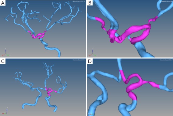
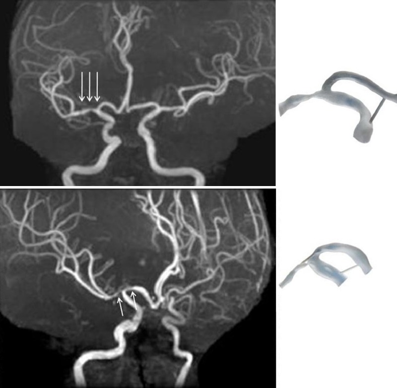
Methods: Three dimensional magnetic resonance angiography (MRA) was performed on two patients with middle cerebral artery (MCA) stenosis. Using scale-adaptive vascular modeling, 3D vascular models were constructed from the MRA source images. The magnified (ten times) regions of interest (ROI) of the stenotic segments were selected and fabricated by a 3D printer with a resolution of 30 µm. A survey to 8 clinicians was performed to evaluate the accuracy of 3D printing results as compared with MRA findings (4 grades, grade 1: consistent with MRA and provide additional visual information; grade 2: consistent with MRA; grade 3: not consistent with MRA; grade 4: not consistent with MRA and provide probable misleading information). If a 3D printing vessel segment was ideally matched to the MRA findings (grade 2 or 1), a successful 3D printing was defined.
Results: Seven responders marked "grade 1" to 3D printing results, while one marked "grade 4". Therefore, 87.5% of the clinicians considered the 3D printing were successful.
Conclusions: Our pilot study confirms the feasibility of using 3D printing technique in the research field of intracranial artery diseases. Further investigations are warranted to optimize this technique and translate it into clinical practice.
Keywords: Three dimensional printing (3D printing); intracranial stenosis; magnetic resonance angiography (MRA); stroke.
Figures
References
-
- Gerstle TL, Ibrahim AM, Kim PS, et al. A plastic surgery application in evolution: three-dimensional printing. Plast Reconstr Surg 2014;133:446-51. - PubMed
-
- Mashiko T, Otani K, Kawano R, et al. Development of Three-Dimensional Hollow Elastic Model for Cerebral Aneurysm Clipping Simulation Enabling Rapid and Low Cost Prototyping. World Neurosurg 2013. [Epub ahead of print]. - PubMed
-
- Schubert C, van Langeveld MC, Donoso LA. Innovations in 3D printing: a 3D overview from optics to organs. Br J Ophthalmol 2014;98:159-61. - PubMed
LinkOut - more resources
Full Text Sources