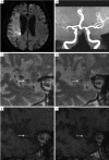
Luminal thrombosis in middle cerebral artery occlusions: a high-resolution MRI study
- PMID: 25333050
- PMCID: PMC4200643
- DOI: 10.3978/j.issn.2305-5839.2014.07.10
Luminal thrombosis in middle cerebral artery occlusions: a high-resolution MRI study
Abstract
Background and purpose: High signals within occluded vessels on T1-weighted fat-suppressed images (HST1) are highly suggestive of luminal thrombosis. We sought to investigate the feasibility of in vivo identification of luminal thrombosis in middle cerebral artery (MCA) occlusions using high-resolution magnetic resonance imaging (HR-MRI).
Methods: We retrospectively reviewed the HR-MRI data of 25 patients with unilateral symptomatic MCA occlusion. HST1 were defined as an area of high signal within the cross-section of occluded MCA, the intensity of which was >150% of the signal of adjacent muscles. The prevalence of HST1 and their relationship to infarct sizes and infarct patterns were analyzed.
Results: The average time from stroke onset to HR-MRI examination was 9±6 days. There were 18 (72%) occluded vessels with HST1 on HR-MRI. HST1 were observed in 5/7 patients with a large territory infarct (≥1/3 MCA distribution) and 13/18 patients without (P=0.37). In the patients with non-large territory infarcts, the presence of HST1 was similar in those with and without border zone infarcts (9/13 vs. 4/5, P=0.42).
Conclusions: It's feasible to in vivo identify luminal thrombosis in occluded MCA. HR-MRI is a potentially powerful tool for investigating the mechanisms of stroke due to MCA occlusions.
Keywords: Middle cerebral artery occlusion (MCA occlusion); cerebral infarct; magnetic resonance imaging; thrombosis.
Figures


References
-
- Klein IF, Lavallée PC, Schouman-Claeys E, et al. High-resolution MRI identifies basilar artery plaques in paramedian pontine infarct. Neurology 2005;64:551-2. - PubMed
-
- Moody AR, Murphy RE, Morgan PS, et al. Characterization of complicated carotid plaque with magnetic resonance direct thrombus imaging in patients with cerebral ischemia. Circulation 2003;107:3047-52. - PubMed
-
- Xu WH, Li ML, Gao S, et al. In vivo high-resolution MR imaging of symptomatic and asymptomatic middle cerebral artery atherosclerotic stenosis. Atherosclerosis 2010;212:507-11. - PubMed
-
- Jansen CH, Perera D, Makowski MR, et al. Detection of intracoronary thrombus by magnetic resonance imaging in patients with acute myocardial infarction. Circulation 2011;124:416-24. - PubMed
LinkOut - more resources
Full Text Sources
