

Global expression profile in low grade meningiomas and schwannomas shows upregulation of PDGFD, CDH1 and SLIT2 compared to their healthy tissue
- PMID: 25333347
- PMCID: PMC4240498
- DOI: 10.3892/or.2014.3526
Global expression profile in low grade meningiomas and schwannomas shows upregulation of PDGFD, CDH1 and SLIT2 compared to their healthy tissue
Abstract
Schwannomas and grade I meningiomas are non‑metastatic neoplasms that share the common mutation of gene NF2. They usually appear in neurofibromatosis type 2 patients. Currently, there is no drug treatment available for both tumors, thus the use of wide expression technologies is crucial to identify therapeutic targets. Affymetrix Human Gene 1.0 ST was used to test global gene expression in 22 meningiomas, 31 schwannomas and, as non-tumoral controls, 3 healthy meningeal tissues, 8 non-tumoral nerves and 1 primary Schwann cell culture. A non-stringent P-value cut-off and fold change were used to establish deregulated genes. We identified a subset of genes that were upregulated in meningiomas and schwannomas when compared to their respectively healthy tissues, including PDGFD, CDH1 and SLIT2. Thus, these genes should be thoroughly studied as targets in a possible combined treatment.
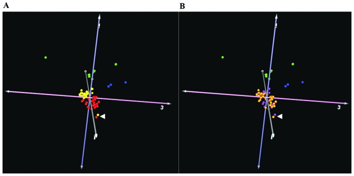
Figures
References
-
- Louis DN, Ohgaki H, Wiestler OD, Cavenee WK, editors. WHO Classification of Tumors of the Central Nervous System. IARC Press; Lyon: 2007. - PubMed
-
- Zankl H, Zang KD. Cytological and cytogenetical studies on brain tumors. 4. Identification of the missing G chromosome in human meningiomas as no. 22 by fluorescence technique. Humangenetik. 1972;14:167–169. - PubMed
-
- Rey JA, Bello MJ, De Campos JM, Kusak ME, Moreno S. Cytogenetic analysis in human neurinomas. Cancer Genet Cytogenet. 1987;28:187–188. - PubMed
-
- Hadfield KD, Smith MJ, Urquhart JE, Wallace AJ, Bowers NL, King AT, Rutherford SA, Trump D, Newman WG, Evans DG. Rates of loss of heterozygosity and mitotic recombination in NF2 schwannomas, sporadic vestibular schwannomas and schwanno-matosis schwannomas. Oncogene. 2010;29:6216–6221. - PubMed
Publication types
MeSH terms
Substances
LinkOut - more resources
Full Text Sources
Other Literature Sources
Molecular Biology Databases
Research Materials
Miscellaneous
