

Superior long-term repopulating capacity of G-CSF+plerixafor-mobilized blood: implications for stem cell gene therapy by studies in the Hbb(th-3) mouse model
- PMID: 25333506
- PMCID: PMC4268579
- DOI: 10.1089/hgtb.2014.005
Superior long-term repopulating capacity of G-CSF+plerixafor-mobilized blood: implications for stem cell gene therapy by studies in the Hbb(th-3) mouse model
Abstract
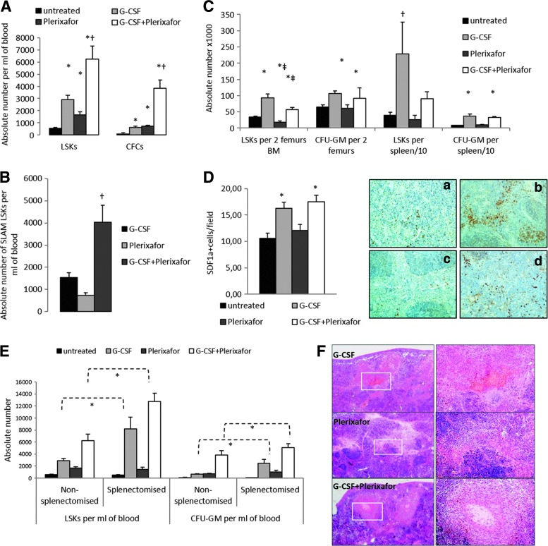
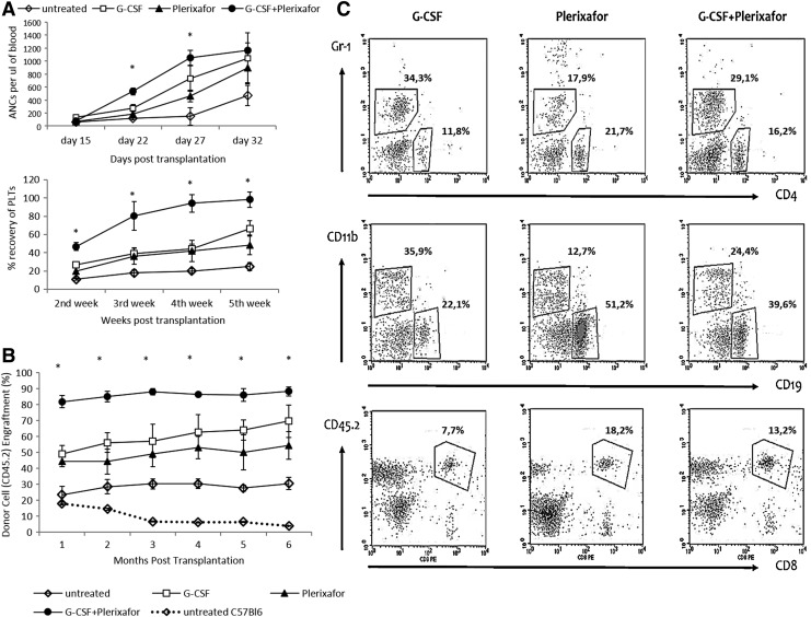
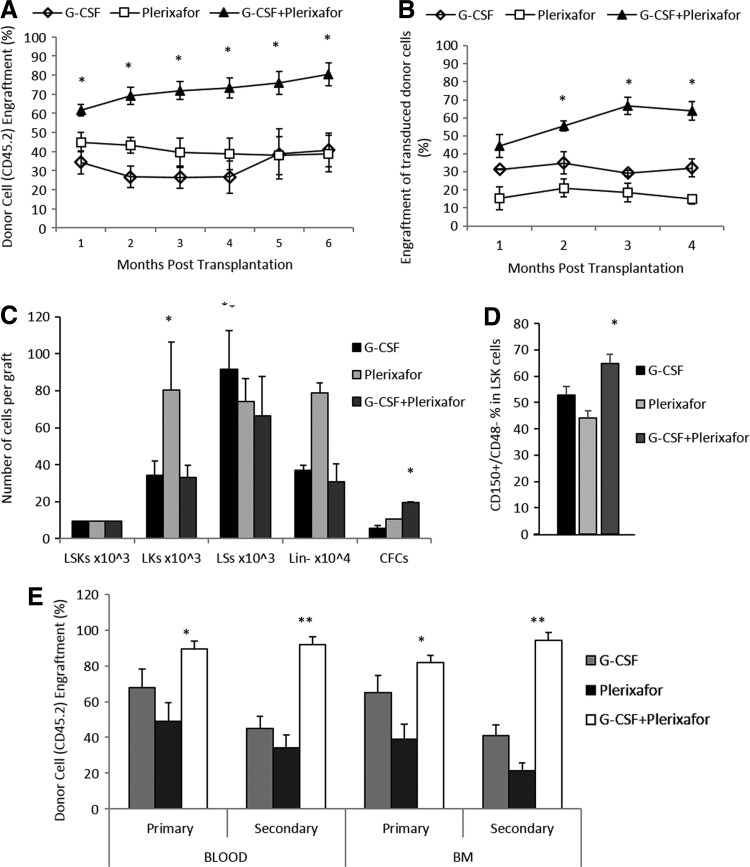
High numbers of genetically modified hematopoietic stem cells (HSCs) equipped with enhanced engrafting potential are required for successful stem cell gene therapy. By using thalassemia as a model, we investigated the functional properties of hematopoietic stem and progenitor cells (HSPCs) from Hbb(th3)/45.2(+) mice after mobilization with G-CSF, plerixafor, or G-CSF+plerixafor and the engraftment kinetics of primed cells after competitive primary and noncompetitive secondary transplantation. G-CSF+plerixafor yielded the highest numbers of HSPCs, while G-CSF+plerixafor-mobilized Hbb(th3)/45.2(+) cells, either unmanipulated or transduced with a reporter vector, achieved faster hematologic reconstitution and higher levels of donor chimerism over all other types of mobilized cells, after competitive transplantation to B6.BoyJ/45.1(+) recipients. The engraftment benefit observed in the G-CSF+plerixafor group was attributed to the more primitive stem cell phenotype of G-CSF+plerixafor-LSK cells, characterized by higher CD150(+)/CD48 expression. Moreover, secondary G-CSF+plerixafor recipients displayed stable or even higher chimerism levels as compared with primary engrafted mice, thus maintaining or further improving engraftment levels over G-CSF- or plerixafor-secondary recipients. Plerixafor-primed cells displayed the lowest competiveness over all other mobilized cells after primary or secondary transplantation, probably because of the higher frequency of more actively proliferating LK cells. Overall, the higher HSC yields, the faster hematological recovery, and the superiority in long-term engraftment indicate G-CSF+plerixafor-mobilized blood as an optimal graft source, not only for thalassemia gene therapy, but also for stem cell gene therapy applications in general.
Figures

Similar articles
-
Plerixafor+G-CSF-mobilized CD34+ cells represent an optimal graft source for thalassemia gene therapy.Blood. 2015 Jul 30;126(5):616-9. doi: 10.1182/blood-2015-03-629618. Epub 2015 Jun 18. Blood. 2015. PMID: 26089395 Free PMC article.
-
Hematopoietic stem cell mobilization for gene therapy: superior mobilization by the combination of granulocyte-colony stimulating factor plus plerixafor in patients with β-thalassemia major.Hum Gene Ther. 2013 Oct;24(10):852-60. doi: 10.1089/hum.2013.163. Hum Gene Ther. 2013. PMID: 24001178 Free PMC article.
-
Accelerated lymphocyte reconstitution and long-term recovery after transplantation of lentiviral-transduced rhesus CD34+ cells mobilized by G-CSF and plerixafor.Exp Hematol. 2011 Jul;39(7):795-805. doi: 10.1016/j.exphem.2011.04.002. Epub 2011 Apr 15. Exp Hematol. 2011. PMID: 21549175 Free PMC article.
-
Plerixafor: A chemokine receptor-4 antagonist for mobilization of hematopoietic stem cells for transplantation after high-dose chemotherapy for non-Hodgkin's lymphoma or multiple myeloma.Clin Ther. 2010 May;32(5):821-43. doi: 10.1016/j.clinthera.2010.05.007. Clin Ther. 2010. PMID: 20685493 Review.
-
Plerixafor injection: a hematopoietic stem cell mobilizer in non-Hodgkin lymphoma and multiple myeloma.Expert Rev Hematol. 2016 Aug;9(8):723-32. doi: 10.1080/17474086.2016.1208082. Epub 2016 Jul 15. Expert Rev Hematol. 2016. PMID: 27355397 Review.
Cited by
-
Mechanisms of Sustained Neutrophilia in Patient WHIM-09, Cured of WHIM Syndrome by Chromothripsis.J Clin Immunol. 2018 Jan;38(1):77-87. doi: 10.1007/s10875-017-0457-8. Epub 2017 Nov 24. J Clin Immunol. 2018. PMID: 29177911 Free PMC article.
-
Plerixafor+G-CSF-mobilized CD34+ cells represent an optimal graft source for thalassemia gene therapy.Blood. 2015 Jul 30;126(5):616-9. doi: 10.1182/blood-2015-03-629618. Epub 2015 Jun 18. Blood. 2015. PMID: 26089395 Free PMC article.
-
Optimizing autologous cell grafts to improve stem cell gene therapy.Exp Hematol. 2016 Jul;44(7):528-39. doi: 10.1016/j.exphem.2016.04.007. Epub 2016 Apr 19. Exp Hematol. 2016. PMID: 27106799 Free PMC article. Review.
-
In vivo HSC transduction in humanized mice mediated by novel capsid-modified HDAd vectors.Mol Ther Methods Clin Dev. 2025 Mar 14;33(2):101448. doi: 10.1016/j.omtm.2025.101448. eCollection 2025 Jun 12. Mol Ther Methods Clin Dev. 2025. PMID: 40231246 Free PMC article.
-
In Vivo Hematopoietic Stem Cell Genome Editing: Perspectives and Limitations.Genes (Basel). 2022 Nov 27;13(12):2222. doi: 10.3390/genes13122222. Genes (Basel). 2022. PMID: 36553489 Free PMC article. Review.
References
-
- Ahmed F., Ings S.J., Pizzey A.R., et al. (2004). Impaired bone marrow homing of cytokine-activated CD34+ cells in the NOD/SCID model. Blood 103, 2079–2087 - PubMed
-
- Aiuti A., Brigida I., Ferrua F., et al. (2009). Hematopoietic stem cell gene therapy for adenosine deaminase deficient-SCID. Immunol. Res. 44, 150–159 - PubMed
-
- Bensinger W., Singer J., Appelbaum F., et al. (1993). Autologous transplantation with peripheral blood mononuclear cells collected after administration of recombinant granulocyte stimulating factor. Blood 81, 3158–3163 - PubMed
-
- Bensinger W.I., Martin P.J., Storer B., et al. (2001). Transplantation of bone marrow as compared with peripheral-blood cells from HLA-identical relatives in patients with hematologic cancers. N. Engl. J. Med. 344, 175–181 - PubMed
Publication types
MeSH terms
Substances
Grants and funding
LinkOut - more resources
Full Text Sources
Other Literature Sources
Research Materials
Miscellaneous
