. 2014 Dec;28(12):2025-37.
doi: 10.1210/me.2014-1206.

A role for site-specific phosphorylation of mouse progesterone receptor at serine 191 in vivo

Affiliations

A role for site-specific phosphorylation of mouse progesterone receptor at serine 191 in vivo

Sandra L Grimm et al. Mol Endocrinol. 2014 Dec.

Abstract

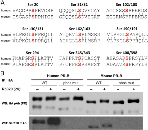
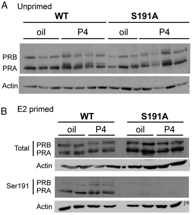
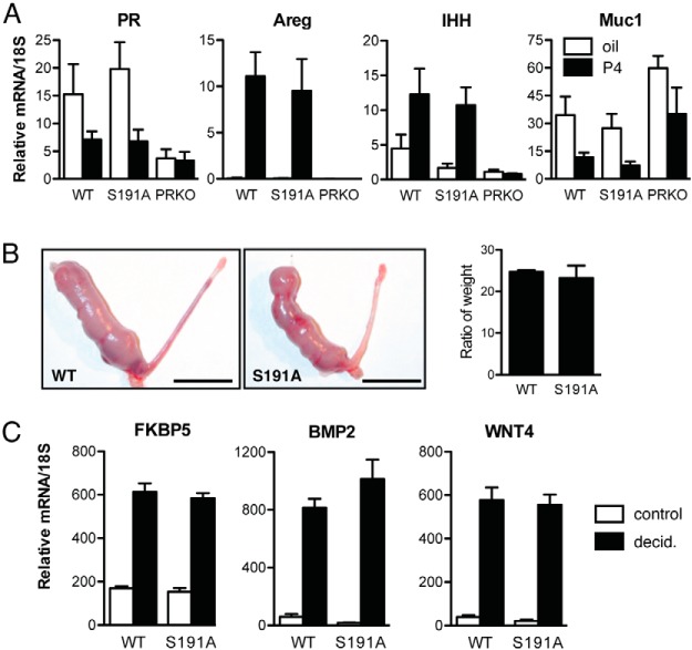
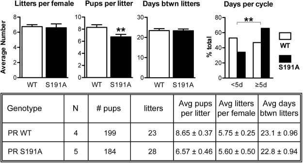
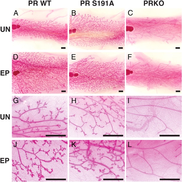
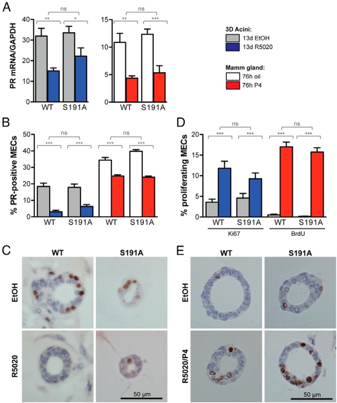
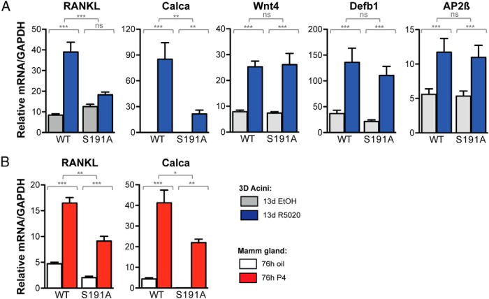
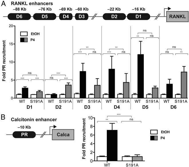
Progesterone receptors (PRs) are phosphorylated on multiple sites, and a variety of roles for phosphorylation have been suggested by cell-based studies. Previous studies using PR-null mice have shown that PR plays an important role in female fertility, regulation of uterine growth, the uterine decidualization response, and proliferation as well as ductal side-branching and alveologenesis in the mammary gland. To study the role of PR phosphorylation in vivo, a mouse was engineered with homozygous replacement of PR with a PR serine-to-alanine mutation at amino acid 191. No overt phenotypes were observed in the mammary glands or uteri of PR S191A treated with progesterone (P4). In contrast, although PR S191A mice were fertile, litters were 19% smaller than wild type and the estrous cycle was lengthened slightly. Moreover, P4-dependent gene regulation in primary mammary epithelial cells (MECs) was altered in a gene-selective manner. MECs derived from wild type and PR S191A mice were grown in a three-dimensional culture. Both formed acinar structures that were morphologically similar, and proliferation was stimulated equally by P4. However, P4 induction of receptor activator of nuclear factor-κB ligand and calcitonin was selectively reduced in S191A cultures. These differences were confirmed in freshly isolated MECs. Chromatin immunoprecipitation analysis showed that the binding of S191A PR to some of the receptor activator of nuclear factor-κB ligand enhancers and a calcitonin enhancer was substantially reduced. Thus, the elimination of a single phosphorylation site is sufficient to modulate PR activity in vivo. PR contains many phosphorylation sites, and the coordinate regulation of multiple sites is a potential mechanism for selective modulation of PR function.


Figures

Figure 1.
Figure 1.
Conserved phosphorylation sites in human and mouse PR. A, A comparison of human and mouse sequences surrounding known phosphorylation sites in human PR. Identity between amino acids shown by bars. B, HEK 293T cells were transduced with human or mouse hemagglutinin (HA)-PR-B lentivirus (WT or phosphomutant) and treated for 2 hours with 0.01% ethanol or 10 nM R5020 before collecting whole-cell extracts. Total PR was immunoprecipitated using an HA polyclonal antibody, run on an SDS-PAGE gel and total PR (HA antibody) and P-Ser191 (Ser190 PR antibody) detected by Western analysis. IP, immunoprecipitation; WB, Western blot.
Figure 2.
Figure 2.
Expression of PR WT and PR S191A protein in mouse uteri is indistinguishable. A, Immunoblotting for total PR in uteri of WT and PR S191A mice pretreated for 6 hours with vehicle (oil) or P4 (1 mg) in non-E2-primed mice. B, Immunoblotting for total (top panel) and phosphorylated (bottom panel) PR (with pS191 specific monoclonal antibody) in uteri of WT and PR S191A mice pretreated for 6 hours with vehicle (oil) or P4 (1 mg) in E2-primed mice.
Figure 3.
Figure 3.
Progesterone-dependent growth and decidualization in uteri of WT and PR S191A C57BL/6 mice do not differ. A, P4-regulated gene expression in uteri from C57BL/6 WT and PR S191A mice. Mice were OVX at 6 weeks of age, rested for 2 weeks, and injected with vehicle (oil) or P4 (1 mg) at 6 hours prior to harvest. RNA was extracted and quantitative PCR was used to analyze expression of selected genes including PR, Areg, Indian hedgehog (Ihh), and mucin-1 (Muc1). Mouse numbers include the following: WT oil, n = 3; WT P4, n = 8; PR S191A oil, n = 7; PR S191A P4, n = 8; PRKO oil, n = 5; and PRKO P4, n = 5. Bars represent SEM. Quantitative PCR values are calculated in relation to PCR standards and normalized to 18S. B, Gross morphology of the uterine decidual response in WT (left panel) and PR S191A (right panel) mice. Graph of the ratio of the traumatized uterine horn weight to the control horn weight of WT (n = 4) vs PR S191A (n = 4) mice, 5 days after the decidual stimulus. Scale bars, 1 cm. C, Expression of select P4-dependent target genes in decidual tissues (FKBP5, BMP2, and Wnt4) in WT and PR S191A mice. WT control (n = 2) and decidual (n = 4) vs PR S191A control (n = 2) and decidual (n = 4) is shown. Control horn, white bars; decidualized horn, black bars. Bars represent SEM. Quantitative PCR values are calculated in relation to PCR standards and normalized to 18S.
Figure 4.
Figure 4.
PR S191A mice have significantly smaller litter sizes and an extended estrous cycle. A 6-month fertility study carried out for the C57BL/6 mice showing the averages (left panel) for the number of litters, pups per litter, and days between litters. Average days between litters for the individual mice between WT (n = 4) and PR S191A mice (n = 5) are shown. A Student's t test was used to determine statistical significance. **, P < .005. A 60-day cyclicity study carried out for the C57BL/6 mice shows the average estrous cycle length for the group (WT, n = 12; PR S191A, n = 11). A Fisher's test was used to determine statistical significance. **, P < .005.
Figure 5.
Figure 5.
Response of mammary gland to prolonged EP treatment. Adult female mice (9–12 wk) were treated with beeswax pellets containing E2 and P4 for a period of 4 weeks as described in Materials and Methods. A–L, Whole-mount staining with Carmine Red at ×10 magnification (A–F) and ×50 magnification (G–L) of inguinal mammary glands from adult (13–14 wk) 129/C57BL/6 mixed WT treated (D and J) and untreated (A and G), PR S191A treated (E and K) and untreated (B and H), and PRKO treated (F and L) and untreated (C and I). Treatment was carried out for 4 weeks. UN, untreated. Animal numbers include the following WT, n = 3 treated, n = 1 untreated; PR S191A, n = 4 treated, n = 2 untreated; PRKO, n = 3 treated, n = 2 untreated; WT on same strain background as PRKO, n = 3 treated, n = 2 untreated. Scale bars, 1 mm. Representative images are shown.
Figure 6.
Figure 6.
Phosphorylation of Ser191 does not alter the pattern of PR expression or P4-induced proliferation of mammary epithelial cells. Primary MECs from WT or PR S191A mice, either acini cultured and treated for 13 days with ethanol vehicle (EtOH) or 100 nM R5020 or glands from mice treated for 76 hours with oil or 1 mg P4, were used to isolate total RNA or were fixed and sectioned to perform IHC. A, Quantitative PCR was performed from triplicate experiments to determine the expression of PR, relative to GAPDH. B, Sections of acini or mammary glands were stained with an anti-PR antibody, and positive cells were quantified relative to total MECs. C, Representative images of PR staining of sectioned WT acini treated with either EtOH or 100 nM R5020 for 13 days. Scale bars, 50 μm. D, Sections of acini or mammary glands were stained with anti-Ki67 or anti-BrdU antibodies, and positive cells were quantified relative to total MECs. E, Representative images of Ki67 staining of sectioned WT acini treated with either ethanol or 100 nM R5020 for 13 days. Scale bars, 50 μm. Statistical significance, determined by a two-way ANOVA, is indicated by one to three asterisks corresponding to P ≤ .05 or P ≤ .01 or P ≤ .001, respectively, or P = ns (not significant).
Figure 7.
Figure 7.
P4 induction of selected target genes is impaired in the mammary epithelial compartment of PR S191A mice. A, Primary MECs from WT or PR S191A mice cultured and treated for 13 days with EtOH or 100 nM R5020 were used to isolate total RNA. Quantitative RT-PCR was performed from triplicate experiments to determine the expression of RANKL, calcitonin (Calca), Wnt4, Defensin-β1, and AP-2β, relative to GAPDH (n = 3 samples in triplicate). B, WT or PR S191A mice were treated in vivo for 76 hours with oil or 1 mg P4. MECs were then harvested, enriched, and used to isolate total RNA. Quantitative RT-PCR was performed to determine the expression of RANKL and calcitonin (Calca), relative to GAPDH. Statistical significance, determined by a two-way ANOVA, is indicated by one to three asterisks corresponding to P ≤ .05 or P ≤ 0.01 or P ≤ 0.001, respectively, or P = ns (not significant).
Figure 8.
Figure 8.
PR S191A mutation alters P4-dependent binding of PR with RANKL or calcitonin enhancers. Recruitment of PR to six distal RANKL enhancers (A) or a calcitonin enhancer (B) was analyzed by a ChIP assay of primary MECs derived from WT or PR S191A mice. Isolated, enriched MECs were treated in vitro for 4.5 hours with vehicle (EtOH) or P4 (100 nM). Immunoprecipitated DNA was amplified by quantitative PCR using primers flanking potential PREs described in the schematic. Amplified ChIP DNA was normalized to the input and displayed as progesterone-induced fold PR recruitment over vehicle, setting the value of vehicle treated primary MECs to 1. Results are mean ± SEM from three independent experiments. Statistical significance, determined by a two-way ANOVA, is indicated by one to three asterisks corresponding to P ≤ .05 or P ≤ .01 or P ≤ .001, respectively, or P = ns (not significant).

References

    1. Hill KK, Roemer SC, Churchill ME, Edwards DP. Structural and functional analysis of domains of the progesterone receptor. Mol Cell Endocrinol. 2012;348(2):418–429. - PMC - PubMed
    1. Ward RD, Weigel NL. Steroid receptor phosphorylation: Assigning function to site-specific phosphorylation. BioFactors. 2009;35(6):528–536. - PMC - PubMed
    1. Trevino LS, Weigel NL. Phosphorylation: a fundamental regulator of steroid receptor action. Trends Endocrinol Metab. 2013;24(10):515–524. - PMC - PubMed
    1. Chen W, Dang T, Blind RD, et al. Glucocorticoid receptor phosphorylation differentially affects target gene expression. Mol Endocrinol. 2008;22(8):1754–1766. - PMC - PubMed
    1. Breitwieser W, Lyons S, Flenniken AM, et al. Feedback regulation of p38 activity via ATF2 is essential for survival of embryonic liver cells. Genes Dev. 2007;21(16):2069–2082. - PMC - PubMed

Publication types