. 2014 Dec;28(12):2049-59.
doi: 10.1210/me.2014-1208.

GnRH evokes localized subplasmalemmal calcium signaling in gonadotropes

Affiliations

GnRH evokes localized subplasmalemmal calcium signaling in gonadotropes

An K Dang et al. Mol Endocrinol. 2014 Dec.

Abstract

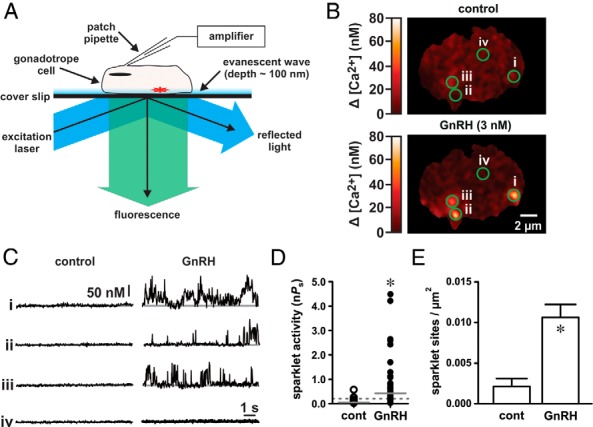
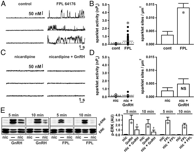
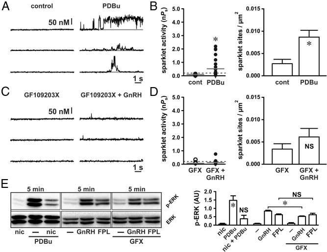
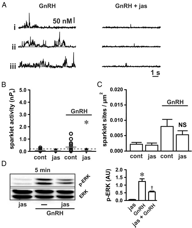
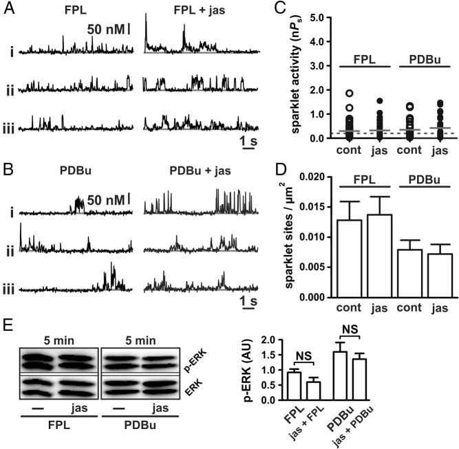
The binding of GnRH to its receptor initiates signaling cascades in gonadotropes, which result in enhanced LH and FSH biosynthesis and secretion. This process is necessary for follicular maturation and ovulation. Calcium influx activates MAPKs, which lead to increased transcription of LH and FSH genes. Previous research suggests that two MAPK signaling pathways, ERK and jun-N-terminal kinase, are activated by either calcium influx through L-type calcium channels or by global calcium signals originating from intracellular stores, respectively. Here we continued this investigation to further elucidate molecular mechanisms transducing GnRH receptor stimulation to ERK activation. Although it is known that GnRH activation of ERK requires calcium influx through L-type calcium channels, direct evidence supporting an underlying local calcium signaling mechanism was lacking. Here we used a combination of electrophysiology and total internal reflection fluorescence microscopy to visualize discrete sites of calcium influx (calcium sparklets) in gonadotrope-derived αT3-1 cells in real time. GnRH increased localized calcium influx and promoted ERK activation. The L-type calcium channel agonist FPL 64176 enhanced calcium sparklets and ERK activation in a manner indistinguishable from GnRH. Conversely, the L-type calcium channel antagonist nicardipine inhibited not only localized calcium sparklets but also ERK activation in response to GnRH. GnRH-dependent stimulation of L-type calcium channels was found to require protein kinase C and a dynamic actin cytoskeleton. Taken together, we provide the first direct evidence for localized L-type calcium channel signaling in αT3-1 cells and demonstrate the utility of our approach for investigating signaling mechanisms and cellular organization in gonadotropes.


Figures

Figure 1.
Figure 1.
GnRH induces localized Ca2+ influx in αT3–1 cells. A, Schematic illustrating the electrophysiological and TIRF imaging method used to visualize Ca2+ influx in αT3–1 cells. B, Representative TIRF images showing localized Ca2+ influx in αT3–1 cells before and after GnRH (3 nM). C, Traces showing the time course of localized Ca2+ influx at the four circled sites before and after GnRH. The scale bar provides reference for changes in [Ca2+]i during the traces (see Materials and Methods). D, Plot of Ca2+ sparklet site activities (nPs; see Materials and Methods) before and after GnRH (n = 32 cells); solid gray lines are the arithmetic means of each group, and dashed lines mark the threshold for high-activity Ca2+ sparklet sites [nPs ≥ .2; (10)]. E, Plot of mean ± SEM Ca2+ sparklet site densities (Ca2+ sparklet sites/μm2) before and after GnRH (n = 32 cells). *, P < .05. cont, control.
Figure 2.
Figure 2.
GnRH-induced Ca2+ sparklets are mediated by L-type Ca2+ channels and promote ERK activation. A and C, Representative traces showing time courses of Ca2+ influx in an αT3–1 cell before and after application of the L-type Ca2+ channel agonist FPL 64176 (500 nM) (A) and GnRH (3 nM) (C) in the presence of the L-type Ca2+ channel antagonist nicardipine (10 μM). B and D, Plots of Ca2+ sparklet site activities (nPs) and mean ± SEM Ca2+ sparklet site densities (Ca2+ sparklet sites per square micrometers) before and after FPL 64176 (n = 12 cells) (B) and GnRH in the presence of nicardipine (n = 7 cells) (D). E, Western blot analysis of ERK activation (as measured by ERK phosphorylation) in αT3–1 cells exposed to GnRH (3 nM) or FPL 64176 (500 nM) in the presence of nicardipine (10 μM) for 5 or 10 minutes (n ≥ 3 independent experiments). *, P < .05 vs nicardipine; †, P < .05 vs nicardipine and P < .05 vs GnRH. cont, control; nic, nicardipine.
Figure 3.
Figure 3.
GnRH-induced L-type Ca2+ channel sparklets and ERK activation require PKC. A and C, Representative traces showing time courses of Ca2+ influx in an αT3–1 cell before and after application of the PKC activator PDBu (50 nM) (A) and GnRH (3 nM) (C) in the presence of the broad-spectrum PKC inhibitor GFX (1 μM). B and D, Plots of Ca2+ sparklet site activities (nPs) and mean ± SEM Ca2+ sparklet site densities (Ca2+ sparklet sites per square micrometer) before and after PDBu (n = 11 cells) (B) and GnRH in the presence of GFX (n = 9 cells) (D). E, Western blot analysis of ERK activation (as measured by ERK phosphorylation) in αT3–1 cells exposed to PDBu (50 nM) or PDBu with nicardipine (1 μM) for 5 minutes and αT3–1 cells exposed to GnRH (3 nM) or FPL 64176 (500 nM) for 5 minutes in the presence or absence of GFX (1 μM; n ≥ 3 independent experiments). *, P < .05. cont, control; nic, nicardipine.
Figure 4.
Figure 4.
Actin stabilization disrupts GnRH-dependent Ca2+ sparklet stimulation. A, Representative traces showing time courses of Ca2+ influx in αT3–1 cells exposed to GnRH (3 nM) in the presence or absence of jasplakinolide (100 nM for 5 min). B and C, Plots of Ca2+ sparklet site activities (nPs) and mean ± SEM Ca2+ sparklet site densities (Ca2+ sparklet sites per square micrometer) for GnRH (3 nM; n = 9 cells in each group) with or without jasplakinolide (100 nM for 5 min). D, Western blot analysis of ERK activation (as measured by ERK phosphorylation) in αT3–1 cells exposed to GnRH (3 nM) with or without jasplakinolide (100 nM for 5 min; n ≥ 3 independent experiments). *, P < .05; †, P < .05 vs jasplakinolide and P < .05 vs GnRH. cont, control; jas, jasplakinolide.
Figure 5.
Figure 5.
Actin stabilization does not disrupt Ca2+ sparklet stimulation by direct activation of L-type Ca2+ channels or PKC. A and B, Representative traces showing time courses of Ca2+ influx in αT3–1 cells exposed to the L-type Ca2+ channel agonist FPL 64176 (500 nM) (A) or the PKC activator PDBu (50 nM) (B) with or without jasplakinolide (100 nM for 5 min). C and D, Plots of Ca2+ sparklet site activities (nPs) and mean ± SEM Ca2+ sparklet site densities (Ca2+ sparklet sites per square micrometer) for FPL 64176 (500 nM; n = 8 cells in each group) and PDBu (50 nM; n = 11 cells in each group) with or without jasplakinolide (100 nM for 5 min). E, Western blot analysis of ERK activation (as measured by ERK phosphorylation) in αT3–1 cells exposed to FPL 64176 (500 nM) and PDBu (50 nM) with or without jasplakinolide (100 nM for 5 min; n ≥ 3 independent experiments). *, P < .05. cont, control; jas, jasplakinolide.

References

    1. Counis R, Laverrière JN, Garrel G, et al. Gonadotropin-releasing hormone and the control of gonadotrope function. Reprod Nutr Dev. 2005;45(3):243–254. - PubMed
    1. Grosse R, Schmid A, Schoneberg T, et al. Gonadotropin-releasing hormone receptor initiates multiple signaling pathways by exclusively coupling to G(q/11) proteins. J Biol Chem. 2000;275(13):9193–9200. - PubMed
    1. Naor Z. Signaling by G-protein-coupled receptor (GPCR): studies on the GnRH receptor. Front Neuroendocrinol. 2009;30(1):10–29. - PubMed
    1. Roberson MS, Misra-Press A, Laurance ME, Stork PJ, Maurer RA. A role for mitogen-activated protein kinase in mediating activation of the glycoprotein hormone alpha-subunit promoter by gonadotropin-releasing hormone. Mol Cell Biol. 1995;15(7):3531–3539. - PMC - PubMed
    1. Mulvaney JM, Zhang T, Fewtrell C, Roberson MS. Calcium influx through L-type channels is required for selective activation of extracellular signal-regulated kinase by gonadotropin-releasing hormone. J Biol Chem. 1999;274(42):29796–29804. - PubMed

Publication types