. 2014 Oct 21;9(10):e111193.
doi: 10.1371/journal.pone.0111193. eCollection 2014.

Expression of the IL-7 receptor alpha-chain is down regulated on the surface of CD4 T-cells by the HIV-1 Tat protein

Affiliations

Expression of the IL-7 receptor alpha-chain is down regulated on the surface of CD4 T-cells by the HIV-1 Tat protein

Denny McLaughlin et al. PLoS One. .

Abstract

HIV infection elicits defects in CD4 T-cell homeostasis in both a quantitative and qualitative manner. Interleukin-7 (IL-7) is essential to T-cell homeostasis and several groups have shown reduced levels of the IL-7 receptor alpha-chain (CD127) on both CD4 and CD8 T-cells in viremic HIV+ patients. We have shown previously that soluble HIV Tat protein specifically down regulates cell surface expression of CD127 on human CD8 T-cells in a paracrine fashion. The effects of Tat on CD127 expression in CD4 T-cells has yet to be described. To explore this effect, CD4 T-cells were isolated from healthy individuals and expression levels of CD127 were examined on cells incubated in media alone or treated with Tat protein. We show here that, similar to CD8 T-cells, the HIV-1 Tat protein specifically down regulates CD127 on primary human CD4 T-cells and directs the receptor to the proteasome for degradation. Down regulation of CD127 in response to Tat was seen on both memory and naive CD4 T-cell subsets and was blocked using either heparin or anti-Tat antibodies. Tat did not induce apoptosis in cultured primary CD4 T-cells over 72 hours as determined by Annexin V and PI staining. Pre-incubation of CD4 T-cells with HIV-1 Tat protein did however reduce the ability of IL-7 to up regulate Bcl-2 expression. Similar to exogenous Tat, endogenously expressed HIV Tat protein also suppressed CD127 expression on primary CD4 T-cells. In view of the important role IL-7 plays in lymphocyte proliferation, homeostasis and survival, down regulation of CD127 by Tat likely plays a central role in immune dysregulation and CD4 T-cell decline. Understanding this effect could lead to new approaches to mitigate the CD4 T-cell loss evident in HIV infection.


Conflict of interest statement

Competing Interests: The authors have declared that no competing interests exist.

Figures

Figure 1
Figure 1. HIV-1 Tat protein down regulates CD127 surface expression on CD4 T-cells.
Purified CD4 T-cells from healthy HIV-negative volunteers were incubated with Tat protein or in medium alone, stained for CD127 and analyzed by flow cytometry. (A) Percent change in CD127+ CD4 T-cells cultured with Tat (10 µg/ml) compared with media controls (n = 17; p<0.01). (B) Change in mean channel fluorescence relative to media controls for the same cultures (p<0.01). (C) Representative flow cytometry histograms from one individual showing progressive down regulation of CD127 surface expression at 48 hours with increasing amounts of Tat protein (black line) compared to medium (grey fill). (D) Composite data (n = 6; p<0.02) showing time and dose response to Tat in percent change in CD127+ CD4 T cells compared to media controls. (E) Purified CD4 and CD8 T-cells from the same healthy HIV-negative volunteers (n = 5) were incubated for up to 72 hours with HIV-1 Tat protein (10 µg/ml) or in medium alone. Percent changes in surface CD127 expression compared to medium controls are shown (p<0.01 for Tat-treated samples; p>0.5 between T-cell subsets). Graphs show mean values ± standard error of the mean (SEM).
Figure 2
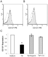
Figure 2. Tat-induced CD127 down regulation is blocked by heparin and anti-Tat antibodies.
Purified Tat protein (2 µg) was pre-incubated with 20 USP heparin or 20 µg anti-Tat polyclonal antibodies (equimolar ratio with Tat) for 30 minutes before being added to CD4 T-cells (n = 4). Representative flow cytometry histograms show CD127 surface expression on CD4 T-cells after 24 hours incubation with (A) 10 µg/ml Tat (grey line), Tat plus heparin (black line) or in medium alone (grey fill); and (B) 10 µg/ml Tat (grey line), Tat plus anti-Tat antibodies (black line) or in medium alone (grey fill). (C) Composite data (n = 5) showing CD127 expression relative to medium controls for Tat, Tat plus heparin, and Tat plus anti-Tat antibodies after 24 hours incubation. * denotes p value <0.05 compared to media. The relative changes are mean values ± SEM.
Figure 3
Figure 3. Tat protein does not affect the viability of purified CD4 T-cells.
Purified CD4 T-cells from healthy HIV-negative volunteers (n = 4) were incubated in medium alone or with Tat protein (10 µg/ml) for up to 72 hours and then stained with propidium iodide or annexin V-FITC and analyzed by flow cytometry. Analyses were performed on the total ungated population as well as on only live cells based on forward and side scatter. (A) Propidium iodide exclusion showed no significant difference in viability between cells cultured in Tat or medium alone. (B) Apoptosis as indicated by positive annexin V staining averaged <10% in the gated population at 72 hours and was no different comparing cells cultured in medium alone or with Tat protein. Camptothecin (10 µM) induces apoptosis and was used as a positive control. * denotes p value <0.05 compared to media. Graphs show mean values ± SEM.
Figure 4
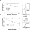
Figure 4. Tat down regulates CD127 surface expression equally on naïve and memory CD4 T-cell subsets.
Purified CD4 T-cells from healthy HIV-negative volunteers (n = 4) were incubated in medium alone or with Tat protein (10 µg/ml) for up to 48 hours. CD127 surface expression was then measured by flow cytometry on naïve CD4 T-cells defined as CD45RA+ and CD62L+, and on memory cells defined as CD45RO+. There was little difference between the subsets (p>0.33). The changes relative to medium controls (p<0.02) are shown (mean values ± SEM). Representative histograms show CD127 expression on indicated populations at 24 hours comparing media (grey fill) to CD4 T-cells treated with Tat protein (black line).
Figure 5
Figure 5. Tat does not affect other CD4 T-cell surface proteins.
Purified CD4 T-cells from healthy HIV-negative volunteers (n = 4) were incubated in medium alone or with Tat protein (10 µg/ml) for up to 48 hours and then analyzed by flow cytometry. (A) Changes in positive staining for the indicated surface proteins relative to medium controls (p>0.09 for all proteins other than CD127 where p<0.04). (B) Change in CD132+ CD4 T-cells relative to medium controls (p>0.12 at 48 hours). Graph represents mean values ± SEM. Representative flow histograms show CD4 T-cells from one individual incubated in media (grey fill) or media plus Tat (black line) at 48 hours.
Figure 6
Figure 6. Tat-induced CD127 down regulation is not due to CD4 T-cell activation.
Purified CD4 T-cells from healthy HIV-negative volunteers (n = 4) were incubated in medium alone or with Tat protein (10 µg/ml) for up to 48 hours and then analyzed by flow cytometry. The changes in surface protein expression relative to medium controls are shown (p>0.19 for all proteins other than CD127 where p<0.04 at 48 hours). Graph represents mean values ± SEM. Representative flow histograms show CD4 T-cells from one individual incubated in media (grey fill) or media plus Tat (black line) at 48 hours.
Figure 7
Figure 7. CD127 surface expression recovers on CD4 T-cells after Tat is removed from the culture medium.
Purified CD4 T-cells from healthy HIV-negative volunteers (n = 4) were incubated in medium alone or with Tat protein (10 µg/ml). At 24 (Tat W 24) and 48 (Tat W 48) hours the cells were washed twice in PBS and resuspended in fresh medium in the absence of Tat protein. Cells were analyzed for CD127 surface expression by flow cytometry at the times indicated. The changes relative to medium controls are shown (mean values ± SEM).
Figure 8
Figure 8. de novo protein synthesis is not required for Tat-induced down regulation of CD127.
Purified CD4 T-cells from healthy HIV-negative volunteers were incubated in medium alone, with cyclohexamide (100 µM; CHX), Tat protein (10 µg/ml) or Tat plus CHX for 24 hours and then analyzed by flow cytometry. (A) Representative histogram showing CD127 surface expression on CD4 T-cells cultured in the presence of CHX alone (black line), Tat alone (dashed line) and CHX plus Tat (grey line) compared to medium control (grey fill). (B) Composite data from n = 4 demonstrating inhibition of protein synthesis with CHX does not prevent Tat-induced CD127 down regulation at the cell surface. CHX alone had no significant effect on CD127 expression (p = 0.07). There was no significant variance in surface CD127 expression between Tat and Tat + CHX treatments (p = 0.38) whereas both treatments showed significant down regulation of CD127 relative to medium (* denotes p<0.009). The percent changes relative to medium controls are shown (mean values ± SEM).
Figure 9
Figure 9. de novo protein synthesis is required for recovery of CD127 surface expression on CD4 T-cells following Tat-induced down regulation.
Purified CD4 T-cells from healthy HIV-negative volunteers (n = 4) were incubated in medium alone or with Tat protein (10 µg/ml). (A) After 24 hours, cells incubated with Tat were washed twice and resuspended in fresh medium alone (Tat W 24) or in medium plus 100 µM CHX (Tat W 24 CHX). Cells were analyzed for CD127 surface expression by flow cytometry at the times indicated. (B) Cells were pre-incubated with and without proteasome inhibitors (5 µM MG132, 10 µM Lactacystin) and lysosome inhibitors (10 µM Leupeptin, 10 µg/ml Pepstatin) for 1 hour followed by incubation with 10 µg/ml Tat protein. After 24 hours, CD127 surface expression was measured by flow cytometry. The changes relative to medium controls are shown (mean values ± SEM). * denotes p value less than 0.05 compared to Tat treated control.
Figure 10
Figure 10. Endogenous production of Tat protein within CD4 T-cells prevents recovery of CD127 on the cell surface.
Isolated CD4 T cells were stimulated with anti-CD3/anti-CD28 beads for 24 hours and then nucleofected with either pcDNA3.1(-) empty vector (mock) or pcDNA3.1(-) expressing Tat. (A) Tat protein expression was confirmed by western blot analysis. Cell lysates were prepared 24 hours following transfection, separated by SDS-PAGE and then analyzed by Western for Tat expression using α-Tat antibodies (clone 1102-Immunodiagnostics). Beta-actin was used as a loading control and purified recombinant Tat (r-Tat) was included to confirm the size and identity of endogenously expressed Tat protein. (B) Surface CD127 expression on CD4 T cells was measured after 24 hours by flow cytometry. Percent change in CD127 expression relative to empty pcDNA3.1(-) control is shown (n = 4; mean values ± SEM). * denotes p value less than 0.05 compared to mock control.
Figure 11
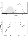
Figure 11. Soluble HIV-1 Tat protein inhibits IL-7 induced Bcl-2 expression in CD4 T-cells.
Purified CD4 T-cells from healthy HIV-negative volunteers were pre-incubated in medium alone or in medium containing Tat protein (10 µg/ml) for 48 hours. Cells were then stimulated with IL-7 (10 ng/ml) for an additional 72 hours and then fixed, stained for Bcl-2 expression and analyzed by flow cytometry. (A) Representative flow cytometry histogram showing Bcl-2 expression in untreated CD4 T-cells (grey fill), following IL-7 stimulation (black line), and following IL-7 stimulation of Tat pre-treated cells (grey line). (B, C) Bcl-2 expression as measured by mean channel fluorescence (MCF) in (B) CD4 T cells treated with IL-7 compared to medium controls, and (C) cells pre-incubated in medium or with Tat prior to IL-7 stimulation (n = 5).

Similar articles

Cited by

References

    1. Embretson J, Zupancic M, Beneke J, Till M, Wolinsky S, et al. (1993) Analysis of human immunodeficiency virus-infected tissues by amplification and in situ hybridization reveals latent and permissive infections at single-cell resolution. Proc Natl Acad Sci U S A 90: 357–361. - PMC - PubMed
    1. Chun TW, Carruth L, Finzi D, Shen X, DiGiuseppe JA, et al. (1997) Quantification of latent tissue reservoirs and total body viral load in HIV-1 infection. Nature 387: 183–188. - PubMed
    1. Clark DR, de Boer RJ, Wolthers KC, Miedema F (1999) T cell dynamics in HIV-1 infection. Adv Immunol 73: 301–327. - PubMed
    1. Hazenberg MD, Hamann D, Schuitemaker H, Miedema F (2000) T cell depletion in HIV-1 infection: how CD4+ T cells go out of stock. Nat Immunol 1: 285–289. - PubMed
    1. McCune JM (2001) The dynamics of CD4+ T-cell depletion in HIV disease. Nature 410: 974–979. - PubMed

Publication types

MeSH terms

Substances