. 2014 Oct 21;9(10):e110688.
doi: 10.1371/journal.pone.0110688. eCollection 2014.

Cytoprotective effects of hydrogen sulfide in novel rat models of non-erosive esophagitis

Affiliations

Cytoprotective effects of hydrogen sulfide in novel rat models of non-erosive esophagitis

Oksana Zayachkivska et al. PLoS One. .

Abstract

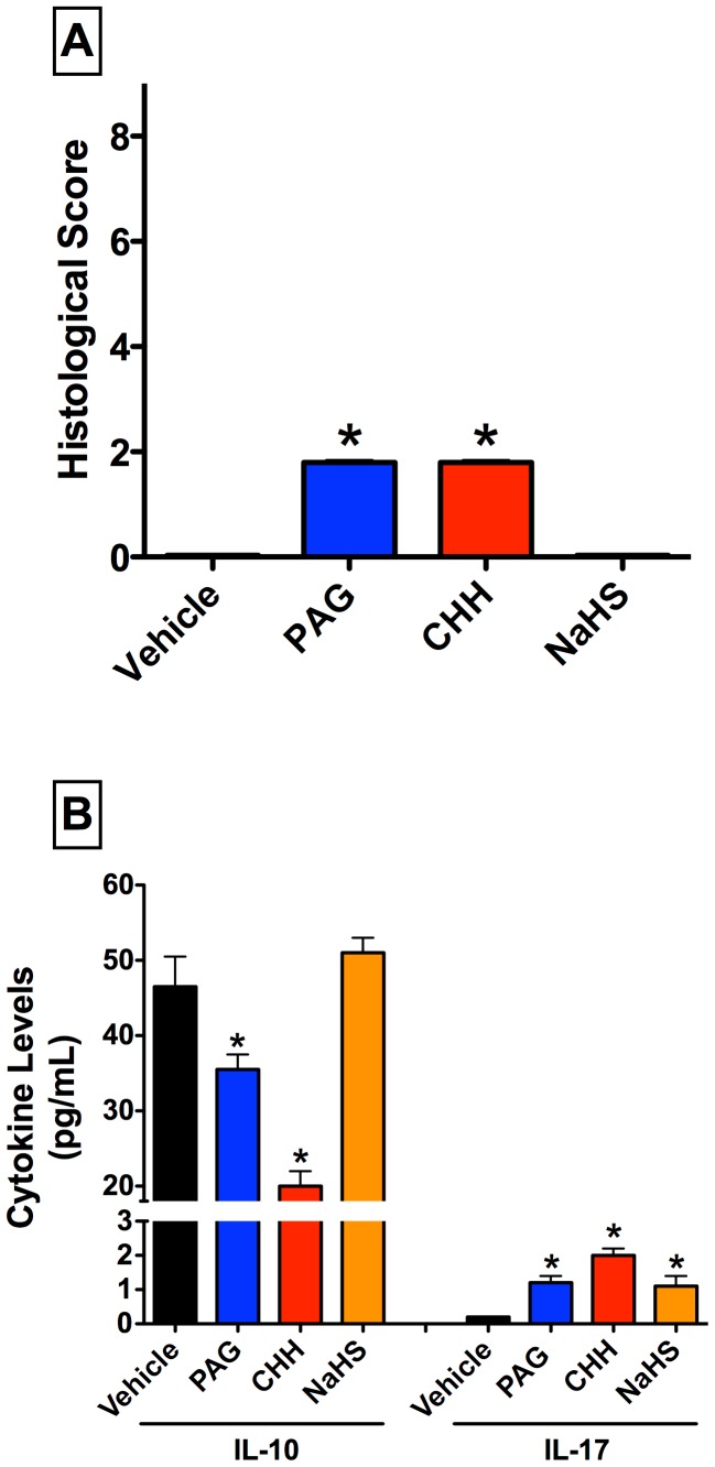
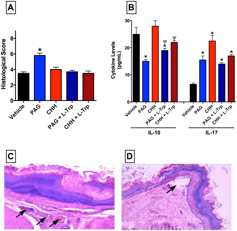
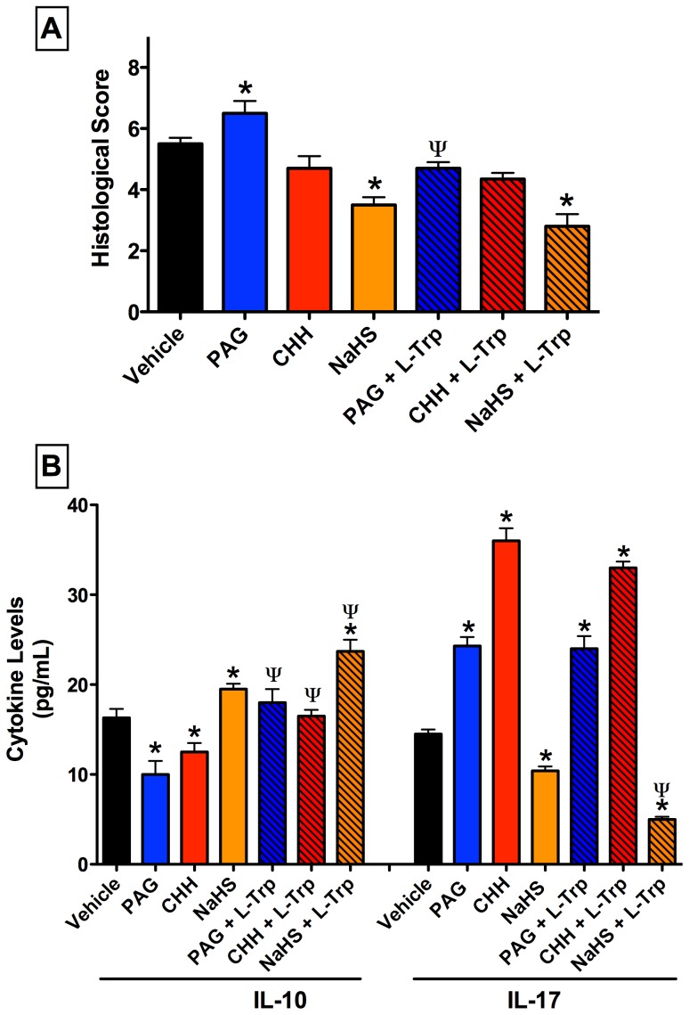
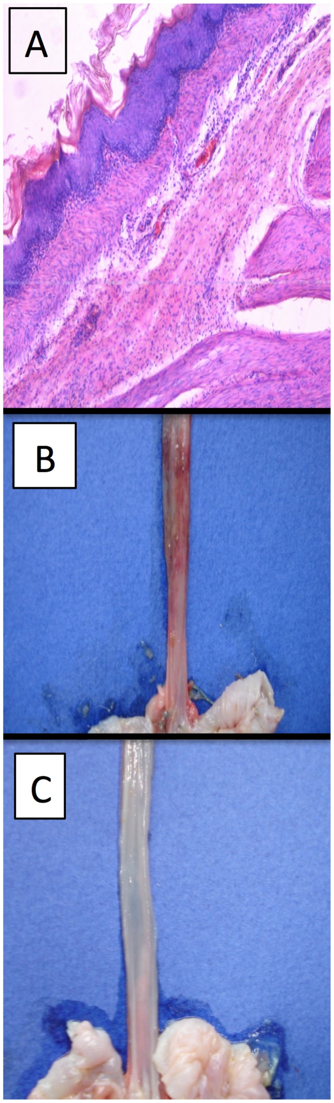
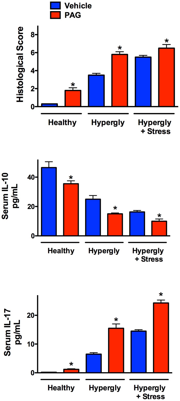
Non-erosive esophagitis is a chronic inflammatory condition of the esophagus and is a form of gastroesophageal reflux disease. There are limited treatment options for non-erosive esophagitis, and it often progresses to Barrett's esophagus and esophageal carcinoma. Hydrogen sulfide has been demonstrated to be a critical mediator of gastric and intestinal mucosal protection and repair. However, roles for H2S in esophageal mucosal defence, inflammation and responses to injury have not been reported. We therefore examined the effects of endogenous and exogenous H2S in rat models of non-erosive esophagitis. Mild- and moderate-severity non-erosive esophagitis was induced in rats through supplementation of drinking water with fructose, plus or minus exposure to water-immersion stress. The effects of inhibitors of H2S synthesis or of an H2S donor on severity of esophagitis was then examined, along with changes in serum levels of a pro- and an anti-inflammatory cytokine (IL-17 and IL-10, respectively). Exposure to water-immersion stress after consumption of the fructose-supplemented water for 28 days resulted in submucosal esophageal edema and neutrophil infiltration and the development of lesions in the muscular lamina and basal cell hyperplasia. Inhibition of H2S synthesis resulted in significant exacerbation of inflammation and injury. Serum levels of IL-17 were significantly elevated, while serum IL-10 levels were reduced. Treatment with an H2S donor significantly reduced the severity of esophageal injury and inflammation and normalized the serum cytokine levels. The rat models used in this study provide novel tools for studying non-erosive esophagitis with a range of severity. H2S contributes significantly to mucosal defence in the esophagus, and H2S donors may have therapeutic value in treating esophageal inflammation and injury.


Conflict of interest statement

Competing Interests: JLW is a director of Antibe Therapeutics Inc., a company developing hydrogen sulfide-releasing anti-inflammatory drugs. This does not alter the authors' adherence to PLOS ONE policies on sharing data and materials.

Figures

Figure 1
Figure 1. Treatment of healthy rats with inhibitors of hydrogen sulfide (H2S) synthesis produces negligible esophageal damage, but significantly alters serum levels of IL-10 and IL-17.
Panel A: Small, but statistically significant changes in the histological score of lower esophageal integrity were observed following administration of either of the inhibitors of H2S synthesis (PAG, L-propargylglycine and CHH, O-carboxymethylhydroxylamine). The changes induced by the H2S inhibitors were limited to mild mucosa inflammation. Administration of the inhibitors of H2S synthesis also resulted in significant decreases in serum IL-10 levels, while the H2S donor, NaHS, had no effect. Small but significant increases in IL-17 levels were observed following administration of PAG, CHH and NaHS. Bars represent the mean ± SEM of at least 6 rats/group. *p<0.05 versus the vehicle-treated group (one-way ANOVA and Neuman-Keuls test).
Figure 2
Figure 2. Administration of an inhibitor of hydrogen sulfide (H2S) synthesis via cystathionine γ-lyase (L-propargylglycine; PAG) exacerbates esophageal injury/inflammation in hyperglycemic rats.
Administration of PAG (25 mg/kg) resulted in a significant increase in the histological score of esophageal injury (panel A). PAG administration also reduced serum IL-10 and increased serum IL-17 levels. The effects of PAG on esophageal damage and serum IL-10 were reversed by co-adminstration of L-tryptophan (L-Trp). An inhibitor of another pathway of H2S synthesis (CHH; O-carboxymethylhydroxylamine; 20 mg/kg) had no effect on esophageal injury, but produced similar changes to serum IL-10 and IL-17 levels as were seen in PAG-treated rats. Panel C shows the irregular hyperemia, stasis (arrows) and perivascular diapedesis that was observed in rats that on the fructose-supplemented drinking water that were treated with PAG. Panel D illustrates that this treatment also resulted in localized detachments of the epithelium from the basement membrane and destructive changes to the epithelial plate (arrow). X200 (hematoxylin and eosin staining). Bars represent the mean ± SEM of at least 6 rats/group. *p<0.05 versus the vehicle-treated group; ψp<0.05 versus the corresponding group not treated with L-tryptophan (one-way ANOVA and Neuman-Keuls test).
Figure 3
Figure 3. Administration of an inhibitor of hydrogen sulfide (H2S) synthesis via cystathionine γ-lyase (L-propargylglycine; PAG) increases esophageal injury/inflammation in hyperglycemic rats subjected to water-immersion stress.
The exacerbation of histological damage by PAG (25 mg/kg) was reversed by co-administration of L-tryptophan (L-Trp). Both inhibitors of H2S synthesis significantly reduced serum IL-10 and increased serum IL-17, and co-administration of L-Trp diminished these effects. The H2S donor, NaHS, significantly increased IL-10 and reduced IL-17, and the combination of NaHS and L-Trp produced significantly greater changes in levels of these two cytokines. Bars represent the mean ± SEM of at least 6 rats/group. *p<0.05 versus the vehicle-treated group; ψp<0.05 versus the corresponding group not treated with L-Trp (one-way ANOVA and Neuman-Keuls test).
Figure 4
Figure 4. Esophageal lesions with signs of nonerosive microscopic esophagitis induced by water-immersion stress after 28 days of consumption of fructose-supplemented drinking water.
Panel A: Pretreatment with an inhibitor of cystathionine γ-lyase (L-propargylglycine; PAG) resulted in extensive esophageal inflammation, with profound submucosal leukocyte infiltration. Panel B: Macroscopic appearance of the lower esophagus of rat treated with PAG, with extensive perivascular hemorrhage. Panel C: Macroscopic appearance of lower esophagus of rat treated with PAG and L-tryptophan, the latter providing a protection against the detrimental effects of PAG.
Figure 5
Figure 5. Summary of some of the key observations in models of mild, moderate and severe esophagitis in rats, and the effects of an inhibitors of hydrogen sulfide synthesis via cystathionine γ-lyase (L-propargylglycine; PAG).
In healthy rats and in the rats with hyperglycemia- or hyperglycemia+stress-induced esophagitis, acute administration of PAG caused a significant exacerbation of esophageal inflammation. Serum levels of IL-10 decreased with the severity of esophagitis, and this was further enhanced in rats treated with PAG. In contrast, serum levels of IL-17 increased sharply in animal with esophagitis, in parallel with the severity of the disease, and administration of PAG caused further increases in all three models. *p<0.05 versus the corresponding vehicle-treated group (Student’s t-test).

References

    1. Konturek SJ, Zayachkivska O, Havryluk XO, Brzozowski T, Sliwowski Z, et al. (2007) Protective influence of melatonin against acute esophageal lesions involves prostaglandins, nitric oxide and sensory nerves. Journal of Physiology and Pharmacology 58: 361–377. - PubMed
    1. Modlin IM, Hunt RH, Malfertheiner P, Moayyedi P, Quigley EM, et al. (2009) Diagnosis and management of non-erosive reflux disease–the Vevey NERD Consensus Group. Digestion 80: 74–88. - PMC - PubMed
    1. Promberger R, Lenglinger J, Riedl O, Seebacher G, Eilenberg W, et al. (2014) Gastro-oesophageal reflux disease in type 2 diabetics: symptom load and pathophysiologic aspects - a retro-pro study. BMC Gastroenterology 14: 132. - PMC - PubMed
    1. Kandulski A, Malfertheiner P (2012) Gastroesophageal reflux disease–from reflux episodes to mucosal inflammation. Nature Reviews Gastroenterology & hepatology 9: 15–22. - PubMed
    1. Kinekawa F, Kubo F, Matsuda K, Kobayashi M, Furuta Y, et al. (2008) Esophageal function worsens with long duration of diabetes. Journal of Gastroenterology 43: 338–344. - PubMed

Publication types

LinkOut - more resources