. 2014 Oct 21;9(10):e110519.
doi: 10.1371/journal.pone.0110519. eCollection 2014.

Infection with Porphyromonas gingivalis exacerbates endothelial injury in obese mice

Affiliations

Infection with Porphyromonas gingivalis exacerbates endothelial injury in obese mice

Min Ao et al. PLoS One. .

Abstract

Background: A number of studies have revealed a link between chronic periodontitis and cardiovascular disease in obese patients. However, there is little information about the influence of periodontitis-associated bacteria, Porphyromonas gingivalis (Pg), on pathogenesis of atherosclerosis in obesity.

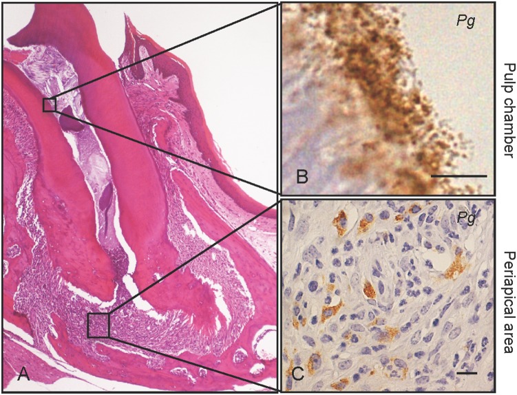
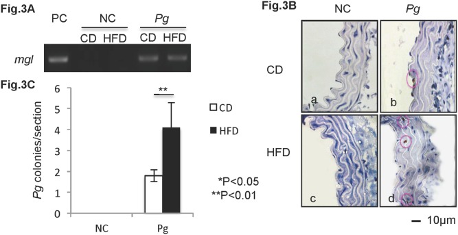
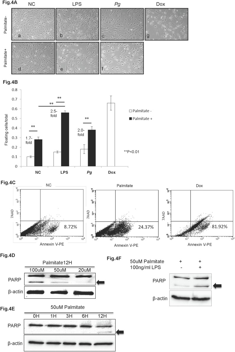
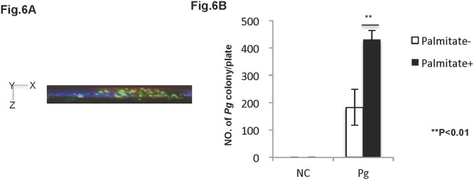
Methods: In vivo experiment: C57BL/6J mice were fed with a high-fat diet (HFD) or normal chow diet (CD), as a control. Pg was infected from the pulp chamber. At 6 weeks post-infection, histological and immunohistochemical analysis of aortal tissues was performed. In vitro experiment: hTERT-immortalized human umbilical vein endothelial cells (HuhT1) were used to assess the effect of Pg/Pg-LPS on free fatty acid (FFA) induced endothelial cells apoptosis and regulation of cytokine gene expression.

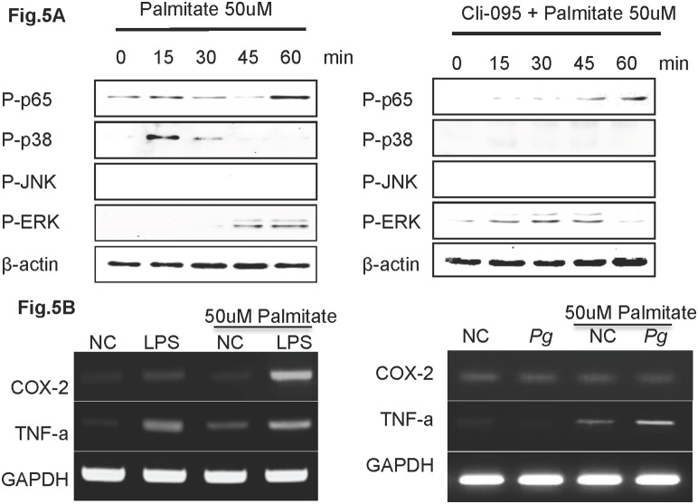
Results: Weaker staining of CD31 and increased numbers of TUNEL positive cells in aortal tissue of HFD mice indicated endothelial injury. Pg infection exacerbated the endothelial injury. Immunohistochemically, Pg was detected deep in the smooth muscle of the aorta, and the number of Pg cells in the aortal wall was higher in HFD mice than in CD mice. Moreover, in vitro, FFA treatment induced apoptosis in HuhT1 cells and exposure to Pg-LPS increased this effect. In addition, Pg and Pg-LPS both attenuated cytokine production in HuhT1 cells stimulated by palmitate.

Conclusions: Dental infection of Pg may contribute to pathogenesis of atherosclerosis by accelerating FFA-induced endothelial injury.


Conflict of interest statement

Competing Interests: The authors have declared that no competing interests exist.

Figures

Figure 1
Figure 1. Dental Pg infection induces periapical granuloma.
Severe pulp necrosis is observed in the first upper molars after six weeks of Pg infection. (A) Periapical granuloma is a representative histological feature of Pg infected molars, and is infiltrated with neutrophils and macrophages. (B) Immunohistochemical staining shows Pg colonies (brown pigment) in the pulp chambers and (C) in neutrophils and macrophages in the periapical area. Scale bar, 10 µm. Pg, Porphyromonas gingivalis. Experiments were performed three times or more with similar results.
Figure 2
Figure 2. Pg exacerbates HFD-induced endothelial injury and apoptosis.
(A) Histological findings in aortal tissue in CD mice (a, b) and in HFD mice (c, d). H&E, scale bar, 100 µm. (B) IHC staining of CD-31 (endothelial cell marker) in aortal tissue in CD mice (a, b) and in HFD mice (c, d). Scale bar, 10 µm. (C) Percentage of aortal surface covered by CD31-positive endothelial cells in the HFD-Pg group is significantly lower than that of the HFD-NC group. Mean ± SD *P<0.05, **P<0.01. (D) TUNEL staining of aortal tissue in CD mice (a, b) and in HFD mice (c, d). Scale bar, 10 µm. (E) Number of TUNEL-positive cells per unit length of aortal surface in HFD-Pg group is significantly higher than that of HFD-NC group. Mean ± SD *P<0.05. CD, chow diet; HFD, high fat diet; NC, negative control; Pg, Porphyromonas gingivalis. Experiments were performed three times or more with similar results.
Figure 3
Figure 3. Dental Pg infection predisposes to increased invasion of injured mouse aortal tissue under HFD conditions.
(A) Expression of the mgl gene of Pg is detected in aortal tissue of infected mice. Genomic DNA of Pg W83 strain is included as a positive control (PC). (B) Immunolocalization of Pg (brown particles) is observed in aortal walls of infected mice. (C) Pg invasion is significantly higher in HFD-Pg group. Mean ± SD *P<0.05, **P<0.01. Scale bar, 10 µm; CD, chow diet; HFD, high fat diet; NC, negative control; Pg, Porphyromonas gingivalis. Experiments were performed three times or more with similar results.
Figure 4
Figure 4. Pg and Pg-LPS increase palmitate-induced apoptosis in HuhT1 cells.
(A, B) Palmitate treatment induces HuhT1 cell detachment, and Pg and/or Pg-LPS treatment further increases detachment. (C) Palmitate stimulation induces cell death by apoptosis that is indicated by increased percentage of Annexin V positive cells; Dox is positive control. (D, E) Palmitate stimulation induces PARP cleavage (arrow, cleaved PARP) in HuhT1 cells in a dose- and time-dependent manner. (F) Pg-LPS accelerates PARP cleavage in palmitate-treated cells. Mean ± SD, **P<0.01. NC, negative control; Pg, Porphyromonas gingivalis; PARP, Poly (ADP-ribose) polymerase; Dox, Doxorubicin. Experiments were performed three times or more with similar results.
Figure 5
Figure 5. Pg-LPS up-regulates COX-2 and TNF-α expression in palmitate treated HuhT1 cells.
(A) Phosphorylation of p65, p38, JNK and ERK was detected by western blotting. Cells were pretreated with Cli-095 6 h before palmitate stimulation. (B) mRNA expression of COX-2 and TNF-α in HuhT1 cells with and without palmitate treatment at 1 h after Pg and/or Pg-LPS stimulation. NC, negative control; Pg, Porphyromonas gingivalis; Cli-095, TLR4 inhibitor. Experiments were performed three times or more with similar results.
Figure 6
Figure 6. Palmitate increases Pg cell invasion in HuhT1 cells.
Antibody protection assay (see Material & Methods) was used to determine Pg invasion in HuhT1 cells. (A) Invaded Pg was analyzed by fluorescent microscopy at magnification of 1000x. Side view (X–Z plane) is shown. Pg (green); DAPI (blue); Phalloidin (red). (B) Pg invasion into HuhT1 cells is increased under palmitate pre-treatment. Mean ± SD, **P<0.01. NC, negative control; Pg Porphyromonas gingivalis. Experiments were performed three times or more with similar results.

Similar articles

Cited by

References

    1. Ross R, Harker L (1976) Hyperlipidemia and atherosclerosis. Science 193(4258): 1094–100. - PubMed
    1. Fuster V, Kelly BB, editors (2010) Promoting Cardiovascular Health in the Developing World: A Critical Challenge to Achieve Global Health. Washington (DC): National Academies Press (US). - PubMed
    1. Libby P (2012) Inflammation in atherosclerosis. Arterioscler Thromb Vasc Biol 32(9): 2045–51 10.1161/ATVBAHA.108.179705 - DOI - PMC - PubMed
    1. Ross R (1999) Atherosclerosis is an inflammatory disease. Am Heart J 138(5 Pt 2): S419–20. - PubMed
    1. Tritto I, Ambrosio G (2004) The multi-faceted behavior of nitric oxide in vascular “inflammation”: catchy terminology or true phenomenon? Cardiovasc Res 63(1): 1–4. - PubMed

Publication types

MeSH terms

LinkOut - more resources