. 2014 Dec 9;53(48):7459-70.
doi: 10.1021/bi501025s. Epub 2014 Nov 24.

Integration host factor assembly at the cohesive end site of the bacteriophage lambda genome: implications for viral DNA packaging and bacterial gene regulation

Affiliations

Integration host factor assembly at the cohesive end site of the bacteriophage lambda genome: implications for viral DNA packaging and bacterial gene regulation

Saurarshi J Sanyal et al. Biochemistry. .

Abstract

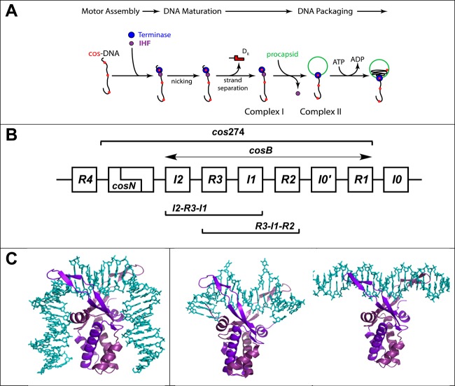
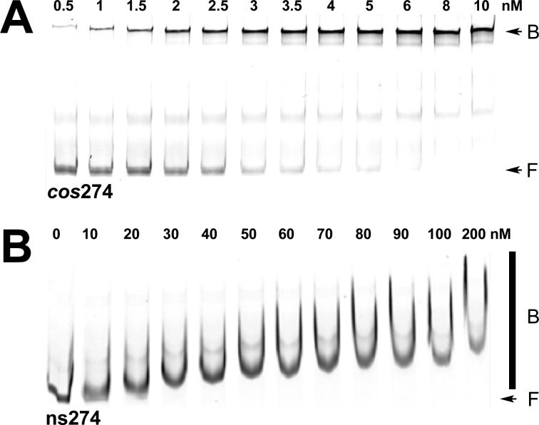
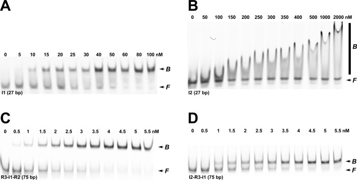
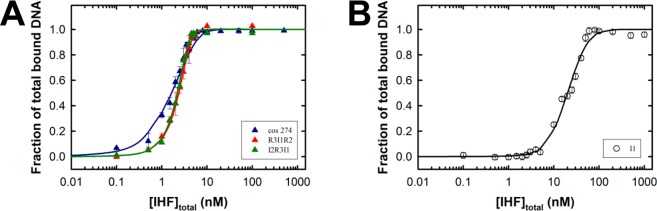
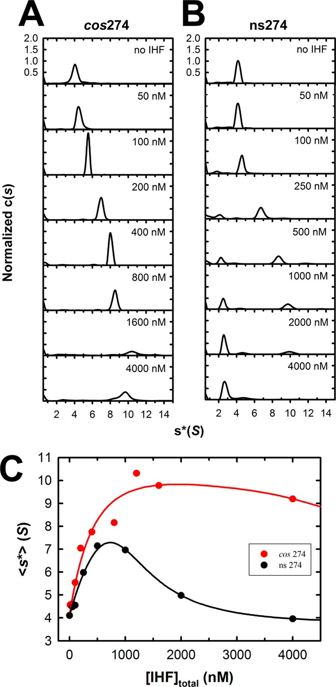
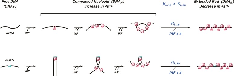
Integration host factor (IHF) is an Escherichia coli protein involved in (i) condensation of the bacterial nucleoid and (ii) regulation of a variety of cellular functions. In its regulatory role, IHF binds to a specific sequence to introduce a strong bend into the DNA; this provides a duplex architecture conducive to the assembly of site-specific nucleoprotein complexes. Alternatively, the protein can bind in a sequence-independent manner that weakly bends and wraps the duplex to promote nucleoid formation. IHF is also required for the development of several viruses, including bacteriophage lambda, where it promotes site-specific assembly of a genome packaging motor required for lytic development. Multiple IHF consensus sequences have been identified within the packaging initiation site (cos), and we here interrogate IHF-cos binding interactions using complementary electrophoretic mobility shift (EMS) and analytical ultracentrifugation (AUC) approaches. IHF recognizes a single consensus sequence within cos (I1) to afford a strongly bent nucleoprotein complex. In contrast, IHF binds weakly but with positive cooperativity to nonspecific DNA to afford an ensemble of complexes with increasing masses and levels of condensation. Global analysis of the EMS and AUC data provides constrained thermodynamic binding constants and nearest neighbor cooperativity factors for binding of IHF to I1 and to nonspecific DNA substrates. At elevated IHF concentrations, the nucleoprotein complexes undergo a transition from a condensed to an extended rodlike conformation; specific binding of IHF to I1 imparts a significant energy barrier to the transition. The results provide insight into how IHF can assemble specific regulatory complexes in the background of extensive nonspecific DNA condensation.


Figures

Figure 1
Figure 1
(A) Assembly of a viral genome maturation and packaging complex at the cos site of the lambda genome. The terminase protomer is composed of one large gpA subunit tightly associated with two smaller gpNu1 subunits. Four protomers and an indeterminate number of IHF dimers cooperatively assemble at a cos sequence of a genome concatemer to engender the packaging motor complex; cos (red dots) represents the junction of two genomes in a concatemer and serves as the packaging initiation site. Terminase and IHF are depicted as blue and purple circles, respectively, for the sake of simplicity. The assembled motor nicks the duplex at cosN to yield the 12-base “sticky” end of the genome (complex I). This intermediate binds a procapsid to yield the functional packaging motor (complex II), which translocates viral DNA into the shell. (B) Detail of the cos region of the lambda genome. The sequence is multipartite consisting of cosN (nicking) and cosB (binding) subsites; cosB extends from I2 to R1 elements. The gpNu1 subunit specifically interacts with the three R elements, and several putative IHF consensus sequences have been identified (I0–I4). The model duplexes used in this study are indicated in the Figure: cos274 (274 bp), [R3-I1-R2] (75 bp), [I2-R3-I1] (75 bp), I1 (27 bp), and R3 (27 bp). (C) Structural models for IHF–DNA nucleoprotein complexes. The left panel shows the crystal structure of IHF bound in a specific complex with the H′ element of attP (PDB entry 1OWF) showing a duplex bend angle of >160°. The DNA binding site size in this complex is ∼34 bp. The middle panel shows the cocrystal structure of Anabaena HU protein bound in a nonspecific complex (PDB entry 1P71) depicting a “weak” (∼105°) bend in the duplex that is found in condensed, nucleoid DNA. The DNA binding site size in this complex is ∼20 bp. The right panel shows the structural model for IHF bound in a nonspecific, linear complex. The model was constructed using MacPymol by manually docking the crystal structure of IHF onto the minimal nonspecific R3 duplex. The DNA binding site size in this complex is ∼8 bp. In all structures, DNA is colored cyan and the α and β subunits of IHF are colored light and dark purple, respectively.
Figure 2
Figure 2
Electrophoretic mobility shift (EMS) studies of binding of IHF to specific (cos274) and nonspecific (ns274) DNA substrates. (A) Representative polyacrylamide gel showing that IHF binds to the specific cos274 substrate to afford a distinct retarded complex. The positions of free (F) and bound (B) DNA complexes are indicated with arrows at the right of the gel image. The band in the middle of the gel represents a contaminant in the IRDye-labeled duplex (Supporting Information). It is unaffected in the titration study and was not considered in the calculation of Fbound. (B) Representative polyacrylamide gel showing that IHF binds to the nonspecific ns274 substrate to afford a concentration-dependent shift and smear on the gel. The positions of free (F) DNA and the bound (B) DNA complexes are indicated at the right of the gel image with an arrow and bar, respectively.
Figure 3
Figure 3
EMS studies of binding of IHF to minimal duplex substrates. (A) Representative polyacrylamide gel showing that IHF binds to the minimal 27 bp I1-specific substrate to afford a distinct retarded complex. We note that upward “smearing” of the retarded band is observed at IHF concentrations of >100 nM (not shown). (B) Representative polyacrylamide gel showing that IHF binds to the minimal 27 bp I2 substrate to afford a smear on the gel. (C) Representative polyacrylamide gel showing that IHF binds to the 75 bp [R3-I1-R2] duplex substrate to afford a distinct retarded complex. (D) Representative polyacrylamide gel showing that IHF binds to the 75 bp [I2-R3-I1] duplex substrate to afford a distinct retarded complex. The positions of free and bound DNA complexes are indicated at the right of each gel image.
Figure 4
Figure 4
Quantitative analysis of EMS binding data. The EMS data (representative data presented in Figures 2 and 3) were converted to fraction bound DNA versus IHF concentration as described in Experimental Procedures. (A) Ensemble of EMS data for binding of IHF to the cos274 (blue), [R3-I1-R2] (red), and [I2-R3-I1] (green) duplexes. Each data point is the average of at least three separate experiments with the standard deviation indicated with error bars. The ensemble of data was simultaneously analyzed according to the nonspecific finite lattice DNA binding model as described in Experimental Procedures. The solid lines represent the best fits of the data, and the derived binding parameters are presented in Table 1. (B) EMS data for binding of IHF to the minimal I1-specific duplex. Each data point is the average of at least three separate experiments with the standard deviation indicated with error bars. The data were analyzed according to the competitive specific/nonspecific finite lattice DNA binding model as described in Experimental Procedures. The solid line represents the best fit of the data, and the derived binding parameters are presented in Table 1.
Figure 5
Figure 5
Interrogation of binding of IHF to minimal substrates using sedimentation velocity analytical ultracentrifugation (SV-AUC). Increasing concentrations of IHF were added to the minimal I1 (specific) and R3 (nonspecific) duplex substrates, and their sedimentation behavior was monitored by SV-AUC as described in Experimental Procedures. The c(s) distribution for each binding experiment was calculated using Sedfit. (A) Normalized c(s) profiles for the specific I1 duplex (27 bp). (B) Normalized c(s) profiles for the nonspecific R3 duplex (27 bp). (C) Weight-average sedimentation coefficients for each of the c(s) distributions shown in panels A (red triangles, I1) and B (black triangles, R3) were calculated using Sedfit and are plotted as a function of IHF concentration. The red dotted line represents the best fit of the I1 binding data to the nonspecific finite lattice DNA binding model, which does not adequately describe the data. The solid lines represent the best fit of simultaneous (global) analysis of the R3 (black) and I1 (red) binding data to (i) nonspecific finite lattice DNA binding and (ii) competitive specific/nonspecific finite lattice DNA models, respectively. The binding parameters derived from global analysis are presented in Table 1.
Figure 6
Figure 6
Interrogation of binding of IHF to full-length cos274 and ns274 duplex substrates using sedimentation velocity analytical ultracentrifugation (SV-AUC). Increasing concentrations of IHF were added to each duplex, and their sedimentation behavior was monitored by SV-AUC as described in Experimental Procedures. The c(s) distribution for each binding experiment were calculated using Sedfit. (A) Normalized c(s) profiles for the specific cos274 duplex. (B) Normalized c(s) profiles for the nonspecific ns274 duplex. (C) Weight-average sedimentation coefficients for each of the c(s) distributions shown in panel A (red circles, cos274) and panel B (black circles, ns274) were calculated using Sedfit and are plotted as a function of IHF concentration. The solid lines represent the best fits of simultaneous (global) analysis of the ensemble of binding data to the DNA unbending model (Scheme 1) as described in Supporting Information. The binding parameters derived from global analysis are presented in Table S2 of the Supporting Information.
Scheme 1
Scheme 1. Model for IHF Nucleoprotein Complexes
Details are provided in the text.

Similar articles

Cited by

References

    1. Azam T. A.; Ishihama A. (1999) Twelve Species of the Nucleoid-associated Protein from Escherichia coli: Sequence Recognition Specificity and DNA Binding Affinity. J. Biol. Chem. 274, 33105–33113. - PubMed
    1. Azam T. A.; Iwata A.; Nishimura A.; Udea S.; Ishihama A. (1999) Growth Phase-Dependent Variation in Protein Composition of the Escherichia coli Nucleoid. J. Bacteriol. 181, 6361–6370. - PMC - PubMed
    1. Drlica K.; Rouviere-Yaniv J. (1987) Histonelike Proteins of Bacteria. Microbiol. Rev. 51, 301–319. - PMC - PubMed
    1. Sarkar T.; Petrov A. S.; Vitko J. R.; Santai C. T.; Harvey S. C.; Mukerji I.; Hud N. V. (2009) Integration Host Factor (IHF) Dictates the Structure of Polyamine-DNA Condensates: Implications for the Role of IHF in the Compaction of Bacterial Chromatin. Biochemistry 48, 667–675. - PubMed
    1. Swinger K. K.; Rice P. A. (2004) IHF and HU: Flexible Architects of Bent DNA. Curr. Opin. Struct. Biol. 14, 28–35. - PubMed

Publication types

MeSH terms

LinkOut - more resources