. 2014 Dec 15;127(Pt 24):5317-30.
doi: 10.1242/jcs.157610. Epub 2014 Oct 21.

Ciliopathy proteins establish a bipartite signaling compartment in a C. elegans thermosensory neuron

Affiliations

Ciliopathy proteins establish a bipartite signaling compartment in a C. elegans thermosensory neuron

Phuong Anh T Nguyen et al. J Cell Sci. .

Abstract

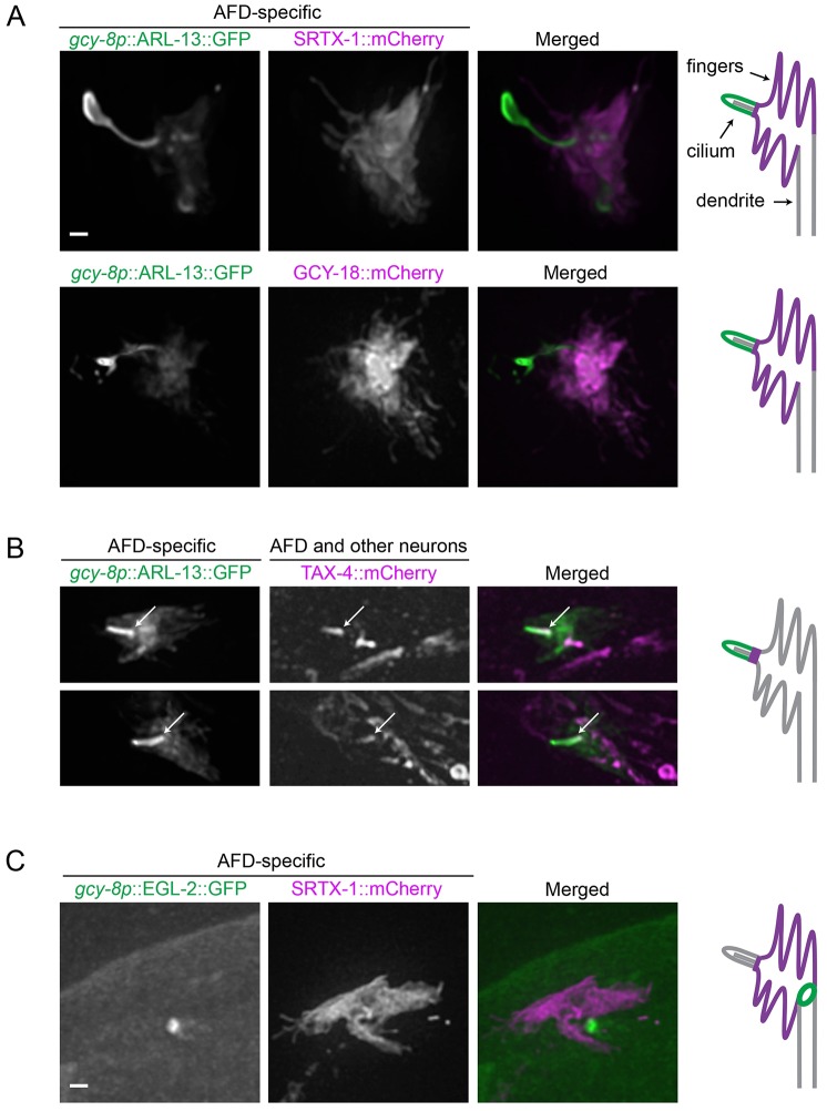
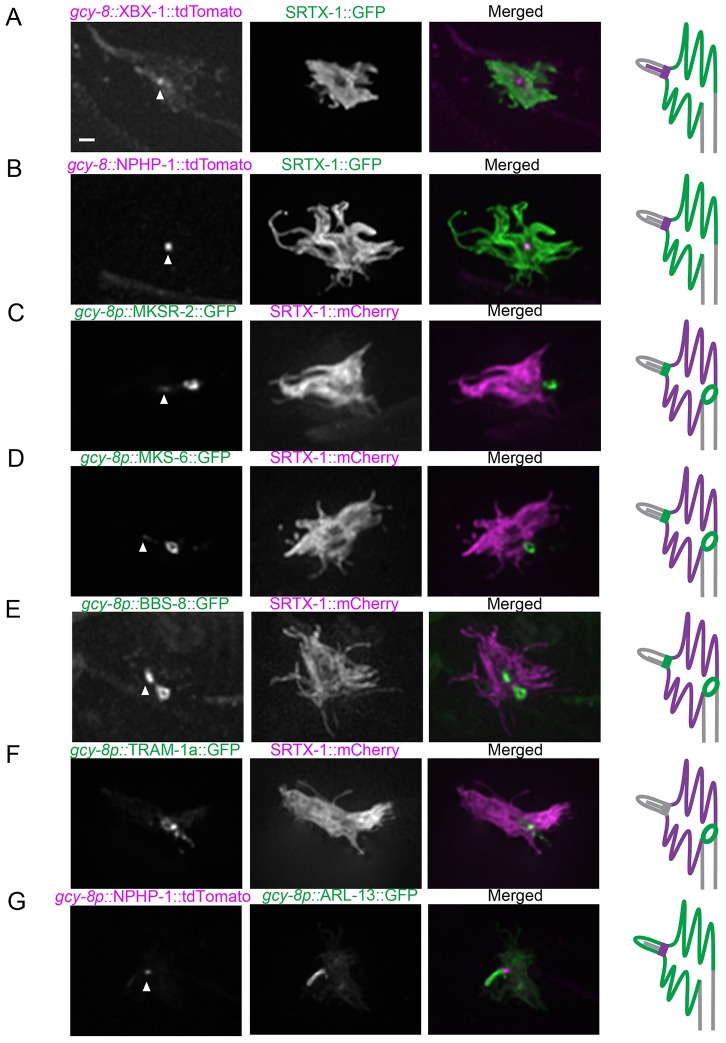
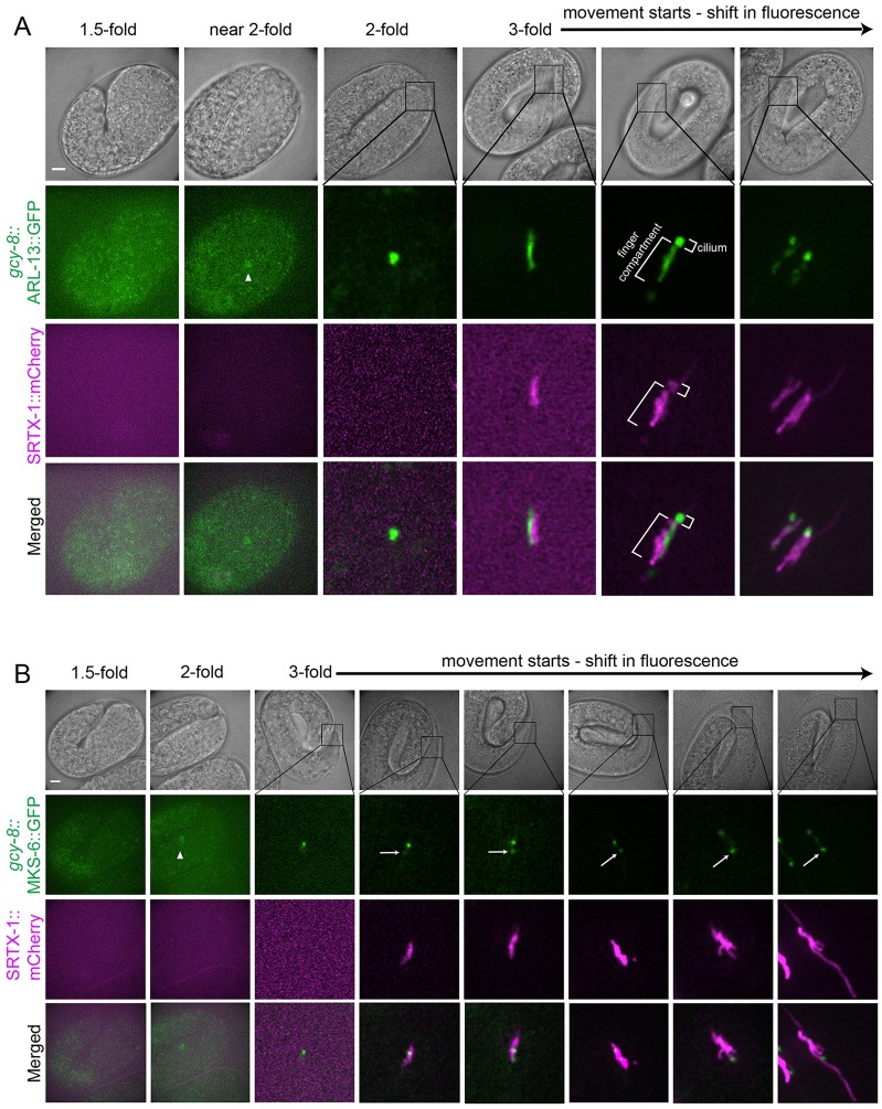
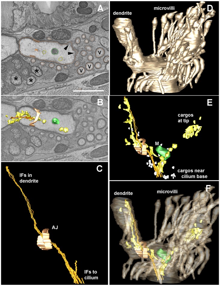
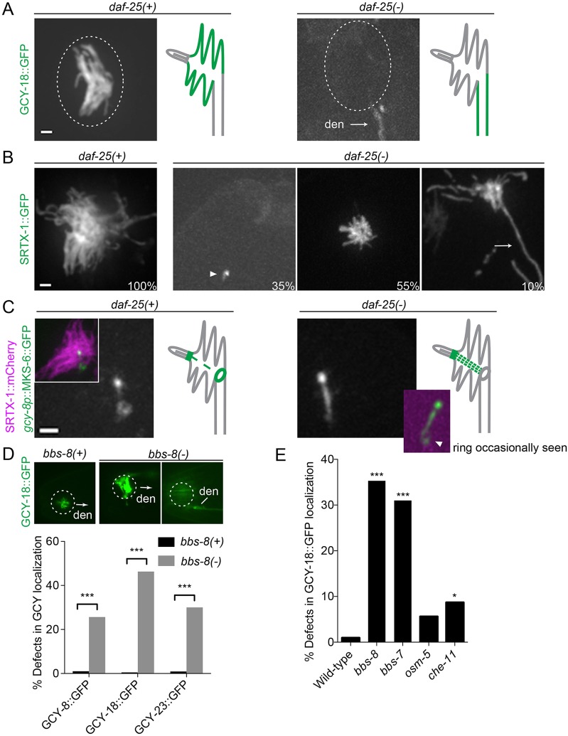
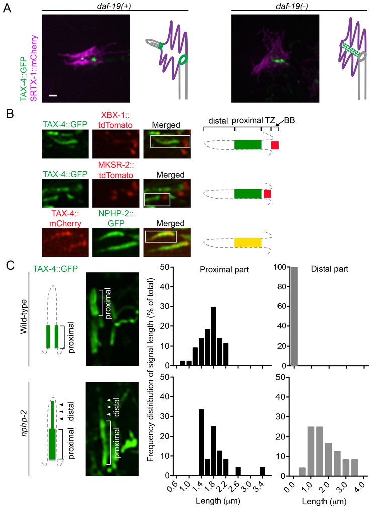
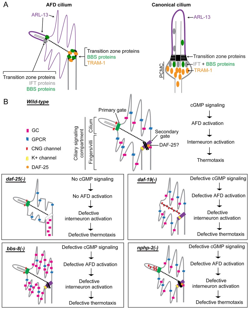
How signaling domains form is an important, yet largely unexplored question. Here, we show that ciliary proteins help establish two contiguous, yet distinct cyclic GMP (cGMP) signaling compartments in Caenorhabditis elegans thermosensory AFD neurons. One compartment, a bona fide cilium, is delineated by proteins associated with Bardet-Biedl syndrome (BBS), Meckel syndrome and nephronophthisis at its base, and requires NPHP-2 (known as inversin in mammals) to anchor a cGMP-gated ion channel within the proximal ciliary region. The other, a subcompartment with profuse microvilli and a different lipid environment, is separated from the dendrite by a cellular junction and requires BBS-8 and DAF-25 (known as Ankmy2 in mammals) for correct localization of guanylyl cyclases needed for thermosensation. Consistent with a requirement for a membrane diffusion barrier at the subcompartment base, we reveal the unexpected presence of ciliary transition zone proteins where no canonical transition zone ultrastructure exists. We propose that differential compartmentalization of signal transduction components by ciliary proteins is important for the functions of ciliated sensory neurons.

Keywords: Compartmentalization; Primary cilia; Sensory neuron; Thermotaxis; Transition zone; cGMP signaling.


Figures

Fig. 1.
Fig. 1.
cGMP signaling components are localized to two distinct compartments in AFD neurons. (A,B) Fluorescently tagged membrane proteins in the cGMP signaling pathway are coexpressed with ARL-13, a ciliary membrane marker, driven by an AFD-specific promoter (gcy-8p). (A) SRTX-1 and GCY-18 are AFD-specific, and present within the finger membrane but not the cilium. (B) TAX-4 is expressed in many ciliated neurons besides AFD neurons, and is localized specifically to the proximal part of the AFD cilium (arrows). Two representative images are shown for TAX-4. (C) EGL-2, an EAG K+ channel, shows ring-like localization pattern at the base of the finger compartment in AFD neurons, as marked by SRTX-1. Schematics of the two compartments and localization of signaling components are depicted on the right of the panels (only six fingers are drawn, there are normally about 60). Scale bars: 1 µm.
Fig. 2.
Fig. 2.
Localization of ciliary and dendritic protein markers uncovers the AFD finger compartment as a cilium-related subcompartment. Fluorescently tagged ciliary proteins were produced specifically in the AFD neurons and co-expressed with SRTX-1, a marker of the AFD finger membrane. The IFT-dynein subunit XBX-1 (A) and transition zone protein NPHP-1 (B) are enriched at the base of the cilium. The transition zone proteins MKSR-2 (C) and MKS-6 (D) are localized at the base of the cilium and are also present as ring structures between the finger and dendritic membranes. (E) BBS-8 is also localized in the cilium and at the ring structure. (F) TRAM-1a, which is normally found at the dendritic tips but not inside cilia, is present at the ring outside of the finger compartment in AFD neurons. (G) Co-expression with ARL-13 confirmed that NPHP-1 signal is at the base of the cilium. Arrowheads indicate the base of the cilium. Scale bar: 1 µm.
Fig. 3.
Fig. 3.
Development of the cilium precedes finger formation. The development of the ciliary and finger compartments in AFD neurons are shown at various stages during embryogenesis. Non-overlapping signals at the 3-fold stage are caused by slight movement of the embryos, which could not be completely eliminated without affecting their development during the experimental time period. (A) Ciliary membrane (marked by ARL-13) appears before the fingers (marked by SRTX-1) are formed. (B) The transition zone protein MKS-6 appears first at the base of the cilium, and then at a more posterior second location (arrows), as the finger compartment (marked by SRTX-1) develops. Arrowheads indicate AFD cell bodies. Scale bars: 5 µm.
Fig. 4.
Fig. 4.
An apical junction separates the finger compartment from the dendritic membrane. (A) The ring-like structure is still present in mutants lacking the ciliary transcription factor DAF-19 or the essential transition zone protein MKS-5. Two neurons are shown for each worm. GFP signals are overexposed to highlight the complete absence of the canonical transition zone in the mutants (arrowheads in wild-type). Overexposure also shows a filamentous structure in wild-type worms decorated with MKS-6 that could be IFs (arrows). Scale bar: 1 µm. (B) In the head, apical junctions (marked by AJM-1) are seen as ring-like structures at the base of cilia (marked by XBX-1) in the bilateral amphid channels (one side is circled). Scale bar: 2 µm. (C) AJM-1 is colocalized with MKS-6 at the ring in AFD neurons. Note the absence of AJM-1 at the canonical transition zone (arrowhead). Scale bar: 0.5 µm.
Fig. 5.
Fig. 5.
Electron tomography reveals the structure of the AFD dendritic ending. An electron tomogram was produced from thick serial sections for a distal portion of the AFD dendrite, in order to highlight details of the microvilli, the apical junction (AJ, pale orange) to the surrounding amphid sheath cell, and the various contents within the finger compartment, which lie along a cluster of IFs that span from the distal dendrite to the base of the cilium. (A) Orthoslice through a portion of the tomogram showing a variety of objects that have been modeled using IMOD to trace their contours, including a mitochondrion (green) and larger vesicles (beige) that might represent smooth ER. Several closely spaced IFs (gold) run as a tight bundle, with these other small objects lying close by. No microtubules were identified within this region. Several amphid channel cilia (asterisks) and several additional microvilli (V) that were not traced are indicated. Arrowheads indicate the base of two traced microvilli; their cytoplasm is locally more electron dense, representing diffuse actin just beneath the plasma membrane. The outline of traced microvilli is shown in the same color as the AFD plasma membrane (beige). Scale bar: 1 µm. (B) A nearby orthoslice shows three-dimensional features of several modeled objects to better display how they fit within the distal dendrite. (C) Side view of modeled region highlights the extension of the IF bundle from inside the distal dendrite, running past the apical junction to enter the finger compartment. This IF bundle continues anteriorward, outside of the tomogram to reach the base of the cilium (not shown), further anterior to the tomogram. (D) Lateral view of the modeled region highlighting the plasma membrane of AFD (in beige), including the distal dendrite and part of the finger compartment. (E) Same model view as C and D, showing the multiple cargoes that lie along the IF bundle. Close to the base of the cilium, many small vesicles (white) cluster near the IF bundle. Smooth ER clusters along the IF bundle both inside the dendrite and within the finger compartment. (F) Same view as panels C, D, E, highlighting the clustering of cargos inside the finger compartment, but with no cargos entering into the surrounding microvilli.
Fig. 6.
Fig. 6.
The daf-25 and bbs-8 mutants show defects in the localization of guanylyl cyclases. (A) Localization of guanylyl cyclases in wild-type and daf-25(-) worms. Guanylyl cyclases, represented here by GCY-18, are absent from AFD fingers of the daf-25 mutant, and are seen along the dendrite (den) instead. Dotted ovals indicate the position of the finger compartment. (B) daf-25(-) worms show defects in finger formation. The numbers are the percentages of worms with each phenotype (n = 100). The arrowhead indicates a complete absence of fingers, and the arrow points to an extra-long finger. (C) The transition zone protein MKS-6 displays an altered localization in daf-25 worms, as it appears stronger at the filamentous structure connecting the cilium and the ring structure (arrowhead indicates the signal occasionally seen in the ring structure in the mutant). Scale bars: 1 µm. (D) Localization of a representative guanylyl cyclase (GCY-18::GFP) in wild-type and bbs-8 worms. GCY-18 is localized specifically in the finger compartment of wild-type AFD neurons, but accumulates in the fingers and along the dendrite of the bbs-8 mutant. In the image on the right for bbs-8(-), the plane was focused on the dendrite to highlight the accumulation there, this neuron still has GFP signal in the finger compartment. Arrows indicate the direction of the dendrite when it is not visible. The graph shows the percentage of AFD neurons with defective localization of each guanylyl cyclase (n>100). (E) Mislocalization of GCY-18 is a prominent phenotype in bbs mutants (bbs-7, bbs-8) but not other mutants that affect IFT (osm-5 and che-11). The bar graph shows the percentage of AFD neurons with defective GCY-18 localization (n>100). *P<0.05; ***P<0.001 (compared with wild-type, χ2 test).
Fig. 7.
Fig. 7.
The daf-19 and nphp-2 mutants show defects in the localization of TAX-4. (A) TAX-4 is mislocalized in the daf-19 mutant. Instead of its wild-type localization at the ciliary base, TAX-4 in the daf-19 mutant is seen in the finger compartment, probably along the IFs. (B) TAX-4 localizes to the inversin compartment of the cilium. In amphid channel cilia, TAX-4 colocalizes with NPHP-2 in the proximal part of the cilium, above the transition zone (TZ, marked by MKSR-2) and the basal body (BB, marked by XBX-1). (C) TAX-4 is mislocalized in amphid cilia of the nphp-2 mutant. Wild-type cilia display discrete localization of TAX-4 at the proximal and not in the distal part of the cilia, whereas nphp-2 cilia show GFP signal leaking into the distal part of cilia. The histograms show the frequency distribution of the signal length at proximal and distal parts in cilia of wild-type (n = 44) and nphp-2 (n = 24) worms. Scale bars: 1 µm.
Fig. 8.
Fig. 8.
Working model of cGMP signaling compartmentalization by ciliary and other proteins at AFD sensory endings. (A) In AFD neurons, ciliary proteins are present in the cilium proper and also found at the base of the finger compartment, establishing it as a cilium-related subcompartment distinct from the periciliary membrane compartment (PCMC) found in other cilia. (B) Summary of signaling compartments in the AFD cilium of wild-type and ciliary mutant worms. DAF-25 might function at the ring structure to regulate the trafficking of signaling proteins into the finger compartment. In the daf-25 mutant, the integrity of this gate and of the fingers is compromised, and guanylyl cyclases (GC) are no longer found in the fingers, so this mutant is expected to have abrogated cGMP signaling. In the bbs-8 mutant, accumulated guanylyl cyclases in the finger compartment could result in a high basal level of cGMP, interfering with thermotransduction. In the nphp-2 mutant, the discrete CNG channel localization is disrupted, which could result in aberrant ion influx and defective neuronal activation. In the daf-19 mutant, the CNG channel is present in the non-permissive membrane environment, or becomes fixed or more abundant along the IFs, and is therefore rendered non-functional. All four mutants are predicted to have altered AFD function and are therefore defective in thermotaxis.

Similar articles

Cited by

References

    1. Adams N. A., Awadein A., Toma H. S. (2007). The retinal ciliopathies. Ophthalmic Genet. 28, 113–125 10.1080/13816810701537424 - DOI - PubMed
    1. Aveldaño M. I., Bazán N. G. (1983). Molecular species of phosphatidylcholine, -ethanolamine, -serine, and -inositol in microsomal and photoreceptor membranes of bovine retina. J. Lipid Res. 24, 620–627. - PubMed
    1. Badano J. L., Mitsuma N., Beales P. L., Katsanis N. (2006). The ciliopathies: an emerging class of human genetic disorders. Annu. Rev. Genomics Hum. Genet. 7, 125–148 10.1146/annurev.genom.7.080505.115610 - DOI - PubMed
    1. Bae Y-K., Qin H., Knobel K. M., Hu J., Rosenbaum J. L., Barr M. M. (2006). General and cell-type specific mechanisms target TRPP2/PKD-2 to cilia. Development 133, 3859–3870 10.1242/dev.02555 - DOI - PubMed
    1. Beales P. L., Elcioglu N., Woolf A. S., Parker D., Flinter F. A. (1999). New criteria for improved diagnosis of Bardet-Biedl syndrome: results of a population survey. J. Med. Genet. 36, 437–446. - PMC - PubMed

Publication types

MeSH terms

LinkOut - more resources