. 2014 Oct 22:5:5288.
doi: 10.1038/ncomms6288.

Dynamic DNA methylation orchestrates cardiomyocyte development, maturation and disease

Affiliations

Dynamic DNA methylation orchestrates cardiomyocyte development, maturation and disease

Ralf Gilsbach et al. Nat Commun. .

Abstract

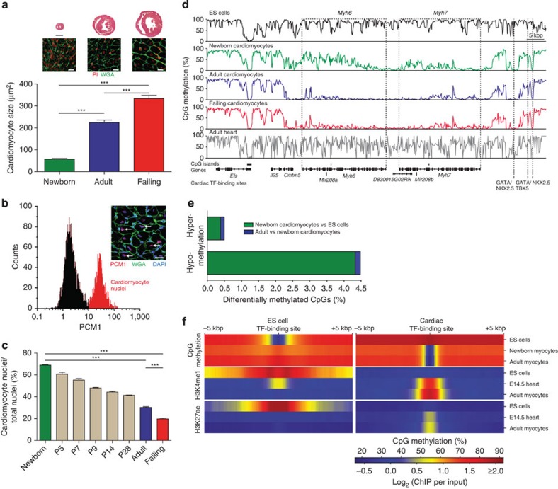
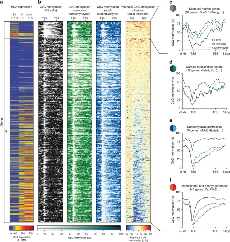
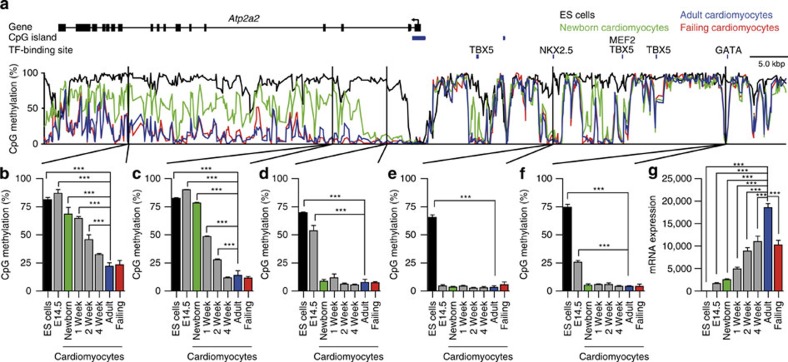
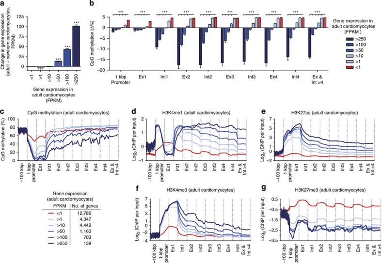
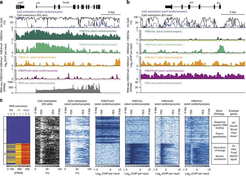
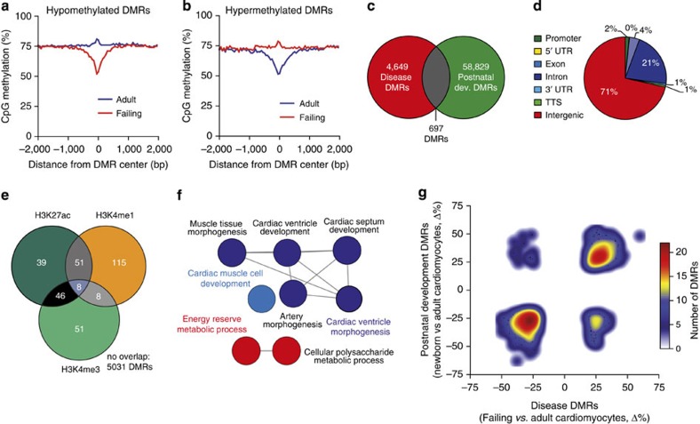
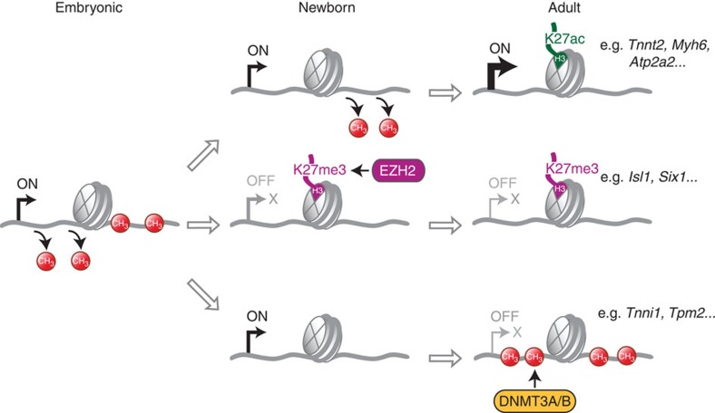
The heart is a highly specialized organ with essential function for the organism throughout life. The significance of DNA methylation in shaping the phenotype of the heart remains only partially known. Here we generate and analyse DNA methylomes from highly purified cardiomyocytes of neonatal, adult healthy and adult failing hearts. We identify large genomic regions that are differentially methylated during cardiomyocyte development and maturation. Demethylation of cardiomyocyte gene bodies correlates strongly with increased gene expression. Silencing of demethylated genes is characterized by the polycomb mark H3K27me3 or by DNA methylation. De novo methylation by DNA methyltransferases 3A/B causes repression of fetal cardiac genes, including essential components of the cardiac sarcomere. Failing cardiomyocytes partially resemble neonatal methylation patterns. This study establishes DNA methylation as a highly dynamic process during postnatal growth of cardiomyocytes and their adaptation to pathological stress in a process tightly linked to gene regulation and activity.


Figures

Figure 1
Figure 1. Analysis of DNA methylation in cardiomyocytes isolated from newborn and adult murine hearts.
(a) Hematoxylin–eosin staining (upper panels; scale bar, 1 mm) of mouse hearts 1 day after birth (newborn), at 8 weeks of age (adult) and 3 weeks after chronic pressure overload in adult mice (failing). WGA-stained ventricular sections (middle panels; scale bar, 20 μm) to determine cardiomyocyte cross-sectional areas (n=10 hearts per group). Nuclei were stained with propidium iodide (PI). (b) Identification of cardiomyocyte nuclei by PCM1 immunostaining in adult mouse cardiac tissue (insert, arrows; scale bar, 20 μm) and purification of cardiomyocyte nuclei (red) by flow cytometry (histogram). (c) Percentage of cardiomyocyte nuclei in ventricular biopsies identified by PCM1 flow cytometry in newborn, adult healthy and failing hearts and at postnatal days 5–28 (n=3–6 hearts per group). (d) IGV (integrative genomics viewer) traces of CpG methylation of the myosin-6 (Myh6) and myosin-7 (Myh7) gene region in ES cells, newborn, adult and failing cardiomyocytes and in murine heart tissue. (e) Differentially methylated CpGs during fetal and postnatal development in percent of assessed CpGs. (f) Density of CpG methylation, histone H3K4me1 and H3K27ac at transcription factor (TF)-binding sites (±5 kbp). Data are shown as mean±s.e.m., ***P<0.001, analysis of variance, Bonferroni post hoc test.
Figure 2
Figure 2. Differentially methylated gene bodies in neonatal and adult cardiomyocytes versus ES cells.
(a,b) Heat maps of gene expression (a) and CpG methylation (b) of gene bodies and flanking regions (±5 kbp) with differential methylation in neonatal and adult cardiomyocytes versus ES cells. Group I, genes with hypermethylated gene bodies in adult cardiomyocytes versus ES cells; group II, genes with hypomethylated gene bodies. (cf) CpG methylation profiles of grouped genes with selected biological function and flanking regions (±5 kbp). Coloured circles refer to Supplementary Figs 8 and 9. TSS, transcription start site; TES, transcription end site.
Figure 3
Figure 3. CpG methylation and gene expression of the Atp2a2 gene.
(af) Gene body demethylation of the Atp2a2 gene and transcription factor-associated DMRs were assessed during fetal and postnatal development in purified cardiomyocyte nuclei by pyrosequencing (assays I (b), II (c), III (d), V (e), VI (f); Supplementary Fig. 4a). (g) Gene expression of Atp2a2 during fetal and postnatal development (ag). Data are shown as mean±s.e.m., n=3–9 per group, ***P<0.001 compared with adult, analysis of variance, Bonferroni post hoc test.
Figure 4
Figure 4. Correlation of gene expression, CpG methylation and histone modifications in genic regions.
(a) Gene expression in adult versus newborn cardiomyocytes was classified according to adult expression levels. Data are shown as mean±s.e.m., n=3, ***P<0.001 adult versus newborn expression, analysis of variance (ANOVA), Bonferroni post hoc test. (b) Changes in DNA methylation of promoters, exon and intron regions between adult and neonatal cardiomyocytes according to adult gene expression levels. Data are shown as mean±s.e.m., n=3, ***P<0.001, ANOVA, Bonferroni post hoc test. (cg) Averaged CpG methylation levels and histone enrichments in genic, promoter (1 kbp) and upstream (100 kbp) regions of adult cardiomyocytes. Genes were grouped according to gene expression levels in adult cardiomyocytes. Ex, exon; Int, intron.
Figure 5
Figure 5. Repression of genes by the polycomb mark H3K27me3.
(a,b) IGV (integrative genomics viewer) traces of CpG methylation of the cardiac troponin T type 2 (Tnnt2) gene (a) and the Isl1 gene (b) in adult cardiomyocytes and ES cells and traces of histone modifications and RNA expression. (c) Heat maps of gene expression, CpG methylation and histone modifications of genes demethylated in adult cardiomyocytes as compared with ES cells. Displayed genes (derived from Fig. 2a, group II) had either very low (<1 FPKM, group 1) or high gene expression (>250 FPKM, group 2) in adult cardiomyocytes (Supplementary Data 2). Right panel, enriched gene ontology terms (P<10−15, hypergeometric test, Bonferroni step down correction) and representative genes. TSS, transcription start site; TES, transcription end site; 5 m CpG, GpG methylation.
Figure 6
Figure 6. Repression of genes by de novo DNA methylation.
(a) Inverse correlation of DNA methylation changes in cardiac myocytes and gene expression changes during postnatal development (P<0.0001, linear regression). (b) Schematic view of a cardiac myofilament at birth (left) and in adult heart (right). Sarcomere components, which are differentially methylated in their genes during postnatal development from birth to adulthood, are filled with green or blue colour. Green colour indicates genes that have low levels of CpG methylation at birth and are significantly higher methylated in adult cardiomyocytes. Blue colour indicates genes with lower methylation in adult versus neonatal cardiomyocytes. Values next to sarcomere components indicate the difference in gene expression (exp) and CpG methylation (me) in adult versus newborn cardiomyocytes. (c,d) DNA methylation traces of troponin I genes Tnni1 (c) and Tnni3 (d) in ES cells, newborn and adult cardiomyocytes (upper panels). Time course of genic DNA methylation (left graphs) and mRNA expression (right graphs). Data are shown as mean±s.e.m., n=3–4 per group, *P<0.05, ***P<0.001 compared with adult, analysis of variance, Bonferroni post hoc test; Dnmt3ab−/− versus adult control, Student’s t-test.
Figure 7
Figure 7. Developmental pattern of CpG methylation in cardiomyocytes in experimental murine heart failure.
(a,b) Overlay plot of CpG methylation in DMRs and surrounding regions of cardiomyocytes isolated from healthy or failing adult hearts displaying hypomethylated (a) and hypermethylated (b) DMRs. (c) Venn diagram showing partial overlap (‘common DMRs’) of disease DMRs (differential methylation between failing and healthy cardiomyocytes) and postnatal DMRs (differential methylation between healthy newborn and adult cardiomyocytes). (d) Genome annotation of DNA regions with differential CpG methylation between failing and healthy adult cardiomyocytes. (e) Intersection of disease DMRs with H3K27ac, H3K4me1 and H3K4me3 peaks in adult cardiomyocytes. (f) Functional annotation of common DMRs in postnatal development and disease (hypergeometric test, Bonferroni step down correction, P value per GO term <0.02). (g) Density plot of common DMRs showing direction of methylation changes in postnatal development and failure. TTS, transcription termination site (region −100 bp to+1 kbp); UTR, untranslated region.
Figure 8
Figure 8. Dynamics of DNA methylation and histone modification in genes bodies of cardiomyocyte genes during development.
Gene bodies of cardiomyocyte genes are sequentially demethylated from embryonic stages (left part, E14.5, data from Fig. 3) until newborn (middle part) and adult stages (right part). The degree of gene body demethylation correlates with histone H3K27ac marks and high expression levels of cardiomyocyte genes (upper trace, data from Figs 1, 3, 5 and 6; Supplementary Fig. 4). Demethylated genes, which are transiently expressed during embryonic development, can be repressed by EZH2-mediated H3K27 trimethylation (middle trace, data from Fig. 5; Supplementary Fig. 12). Demethylated genes may also be repressed by de novo methylation, which is mediated by DNMT3A/B (lower trace, data from Fig. 6; Supplementary Fig. 14).

References

    1. Bergmann O. et al. Evidence for cardiomyocyte renewal in humans. Science 324, 98–102 (2009). - PMC - PubMed
    1. Senyo S. E. et al. Mammalian heart renewal by pre-existing cardiomyocytes. Nature 493, 433–436 (2013). - PMC - PubMed
    1. Taegtmeyer H., Sen S. & Vela D. Return to the fetal gene program: a suggested metabolic link to gene expression in the heart. Ann. NY Acad. Sci. 1188, 191–198 (2010). - PMC - PubMed
    1. Siedner S. et al. Developmental changes in contractility and sarcomeric proteins from the early embryonic to the adult stage in the mouse heart. J. Physiol. 548, 493–505 (2003). - PMC - PubMed
    1. Krüger M., Kohl T. & Linke W. A. Developmental changes in passive stiffness and myofilament ca2+ sensitivity due to titin and troponin-i isoform switching are not critically triggered by birth. Am. J. Physiol. Heart Circ. Physiol. 291, H496–H506 (2006). - PubMed

Publication types

MeSH terms