

Effects of polycaprolactone-based scaffolds on the blood-brain barrier and cerebral inflammation
- PMID: 25335965
- PMCID: PMC4333319
- DOI: 10.1089/ten.TEA.2013.0779
Effects of polycaprolactone-based scaffolds on the blood-brain barrier and cerebral inflammation
Abstract
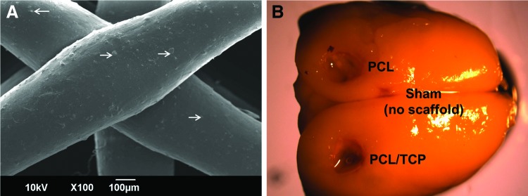
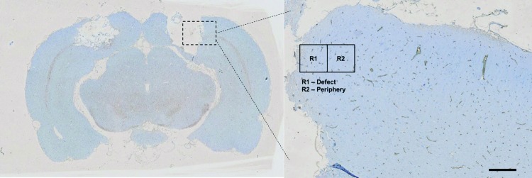
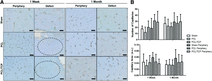
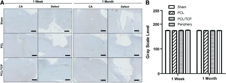
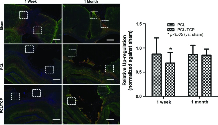
Severe pathoanatomical and mechanical injuries compromise patient recovery and survival following penetrating brain injury (PBI). The realization that the blood-brain barrier (BBB) plays a major role in dictating post-PBI events has led to rising interests in possible therapeutic interventions through the BBB. Recently, the choroid plexus has also been suggested as a potential therapeutic target. The use of biocompatible scaffolds for the delivery of therapeutic agents, but little is known about their interaction with cerebral tissue, which has important clinical implications. Therefore, the authors have sought to investigate the effect of polycaprolactone (PCL) and PCL/tricalcium phosphate (PCL/TCP) scaffolds on the maintenance of BBB phenotype posttraumatic brain injury. Cranial defects of 3 mm depth were created in Sprague Dawley rats, and PCL and PCL/TCP scaffolds were subsequently implanted in predetermined locations for a period of 1 week and 1 month. Higher endothelial barrier antigen (EBA) expressions from PCL-based scaffold groups (p>0.05) were found, suggesting slight advantages over the sham group (no scaffold implantation). PCL/TCP scaffold group also expressed EBA to a higher degree (p>0.05) than PCL scaffolds. Importantly, higher capillary count and area as early as 1 week postimplantation suggested lowered ischemia from the PCL/TCP scaffold group as compared with PCL and sham. Evaluation of interlukin-1β expression suggested that the PCL and PCL/TCP scaffolds did not cause prolonged inflammation. BBB transport selectivity was evaluated by the expression of aquaporin-4 (AQP-4). Attenuated expression of AQP-4 in the PCL/TCP group (p<0.05) suggested that PCL/TCP scaffolds altered BBB selectivity to a lower degree as compared with sham and PCL groups, pointing to potential clinical implications in reducing cerebral edema. Taken together, the responses of PCL-based scaffolds with brain tissue suggested safety, and encourages further preclinical evaluation in PBI management with these scaffolds.
Figures
References
-
- Ding Z., Zhang J., Xu J., Sheng G., and Huang G.Propofol administration modulates aqp-4 expression and brain edema after traumatic brain injury. Cell Biochem Biophys 67,615, 2013 - PubMed
-
- Kazemi H., Hashemi-Fesharaki S., Razaghi S., Najafi M., Kolivand P.H., Kovac S., et al. . Intractable epilepsy and craniocerebral trauma: analysis of 163 patients with blunt and penetrating head injuries sustained in war. Injury 43,2132, 2012 - PubMed
Publication types
MeSH terms
Substances
LinkOut - more resources
Full Text Sources
Other Literature Sources
Medical
