. 2014 Oct 22:4:6716.
doi: 10.1038/srep06716.

Human iPS cell-engineered cardiac tissue sheets with cardiomyocytes and vascular cells for cardiac regeneration

Affiliations

Human iPS cell-engineered cardiac tissue sheets with cardiomyocytes and vascular cells for cardiac regeneration

Hidetoshi Masumoto et al. Sci Rep. .

Abstract

To realize cardiac regeneration using human induced pluripotent stem cells (hiPSCs), strategies for cell preparation, tissue engineering and transplantation must be explored. Here we report a new protocol for the simultaneous induction of cardiomyocytes (CMs) and vascular cells [endothelial cells (ECs)/vascular mural cells (MCs)], and generate entirely hiPSC-engineered cardiovascular cell sheets, which showed advantageous therapeutic effects in infarcted hearts. The protocol adds to a previous differentiation protocol of CMs by using stage-specific supplementation of vascular endothelial cell growth factor for the additional induction of vascular cells. Using this cell sheet technology, we successfully generated physically integrated cardiac tissue sheets (hiPSC-CTSs). HiPSC-CTS transplantation to rat infarcted hearts significantly improved cardiac function. In addition to neovascularization, we confirmed that engrafted human cells mainly consisted of CMs in >40% of transplanted rats four weeks after transplantation. Thus, our HiPSC-CTSs show promise for cardiac regenerative therapy.


Conflict of interest statement

Yes there is potential Competing Interest. T. Okano is a director of the board of CellSeed Inc. T. Okano and T. Shimizu are stake holders of CellSeed Inc. Tokyo Women's Medical University is receiving research fund from CellSeed Inc. J.K. Yamashita is a founder, stake holder, and scientific adviser of iHeart Japan Corporation, and an inventor of pluripotent stem cell-related patents.

Figures

Figure 1
Figure 1. Simultaneous induction of CMs and vascular cells from human iPSCs.
(a) Schematic diagram of cardiovascular cell induction protocols. Defined cardiovascular cell populations (cardiomyocytes [CMs], endothelial cells [ECs] and vascular mural cells [MCs]) are systematically differentiated from human iPSCs. Left panel: a protocol to induce exclusively CMs. Right panel: a modified protocol to induce a variety of cardiovascular cells. (b) Schematic representation of the cardiovascular cell induction protocol. (c) Flow cytometry analysis of cellular components on d15 (n = 13, VEGF 50 ng/ml). hiPSC, human induced pluripotent stem cell; VEGF, vascular endothelial cell growth factor; Dkk1, Dickkopf-related protein 1; MEF-CM, mouse embryonic fibroblast conditioned medium; bFGF, basic fibroblast growth factor; ActA, Activin A; BMP4, bone morphogenetic protein 4; cTnT, cardiac troponin-T; VE-cad, vascular-endothelial cadherin; PDGFRb, platelet-derived growth factor receptor beta.
Figure 2
Figure 2. Generation of hiPSC-cardiac tissue sheets (CTSs).
(a) Schematic diagram of hiPSC-CTS formation. Cardiac cell populations (CMs, ECs, and MCs) are differentiated from hiPSCs, collected, then re-cultured to construct tissue sheets. (b) Macroscopic appearance of hiPSC-CTS. Left panel: 1-ply. Right panel: 3-ply. (c,d) Tissue sheet components. (c) Histological analyses. Upper left panel: Hematoxylin-Eosin (HE) staining. Upper right panel: Sirius-red staining (red) for a cross-section showing intact extracellular matrix. Lower panel: Immunostaining of a sheet for cTnT (CMs, red), and DAPI (cell nuclei, blue). (d) Cellular components analyzed by flow cytometry (n = 13). DAPI, 4, 6 diamidino -2-phenylindole. Scale bars: 1 cm in b, 100 μm in c (lower panel), and 20 μm in c (upper panels).
Figure 3
Figure 3. Echocardiograms of cardiac functions after hiPSC-CTS transplantation to rat MI.
(a) Representative M-mode image of sham-operated rat (left) and CTS transplanted rat (Tx; right). Note that the infarct anterior wall started to contract 2–4 weeks after transplantation in Tx rat. Dotted lines: boundary of anterior or posterior wall of LV. (b) Fractional shortening (FS). (c) Fractional area change (FAC). (d) Systolic thickening. (e) Akinetic length (AL). (f) Diastolic diameter of LV (mm). *P<0.05, **P<0.01 and ***P<0.001 (two-way ANOVA and Tukey's test vs. PreTx). †P<0.05 and ‡P<0.001 (two-way ANOVA). n = 19. PreMI, Pre-induction of MI; PreTx, Pre-transplantation; Tx2w, Tx4w, Tx2mo, 2 weeks, 4 weeks and 2 months after transplantation, respectively; LV, left ventricle.
Figure 4
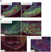
Figure 4. Engraftment of human iPSC-derived cells and neovascularization following hiPSC-CTS transplantation.
(a) Immunostaining with consecutive sections on Tx-d3. Left panel: double staining for vWF (ECs, green) and cTnT (CMs, red), and DAPI. Right panel: double staining for vWF (ECs, green) and HNA (human cell nuclei, red), and DAPI. (b,c) Immunostaining on Tx-d28. (b) Double staining for cTnT (CMs, green) and HNA (human cell nuclei, red), and DAPI. Upper panels: representative staining for engrafted area. Right panel: higher magnification view of left panel (square). Lower panel: quantification of engrafted area. The ratio of the engrafted area (area in the broken magenta line) and the MI area (area in the broken white line) is shown. (c) Double staining for vWF (ECs, green) and HNA (human cell nuclei, red), and DAPI. Tx-d3 and d28: 3 and 28 days after Tx, respectively. Scale bars: 2 mm in b (lower panel), 500 μm in b (upper left panel), 200 μm in a, and 100 μm in b (upper right panel) and c.

References

    1. World Health Organization (WHO), The global burden of disease: 2004 update. http://www.who.int/healthinfo/global_burden_disease/GBD_report_2004updat... (2008) (Date of access: 30/06/2014).
    1. Ford E. S. & Capewell S. Proportion of the Decline in Cardiovascular Mortality Disease due to Prevention Versus Treatment: Public Health Versus Clinical Care. Annu Rev Public Health 32, 5–22 (2011). - PubMed
    1. Committee for Scientific A., Sakata R., Fujii Y. & Kuwano H. Thoracic and cardiovascular surgery in Japan during 2009: annual report by the Japanese Association for Thoracic Surgery. Gen Thorac Cardiovasc Surg 59, 636–667 (2011). - PubMed
    1. Anversa P., Kajstura J., Rota M. & Leri A. Regenerating new heart with stem cells. J Clin Invest 123, 62–70 (2013). - PMC - PubMed
    1. Joggerst S. J. & Hatzopoulos A. K. Stem cell therapy for cardiac repair: benefits and barriers. Expert Rev Mol Med 11, e20 (2009). - PubMed

Publication types