

Amphetamine in adolescence disrupts the development of medial prefrontal cortex dopamine connectivity in a DCC-dependent manner
- PMID: 25336209
- PMCID: PMC4367452
- DOI: 10.1038/npp.2014.287
Amphetamine in adolescence disrupts the development of medial prefrontal cortex dopamine connectivity in a DCC-dependent manner
Abstract
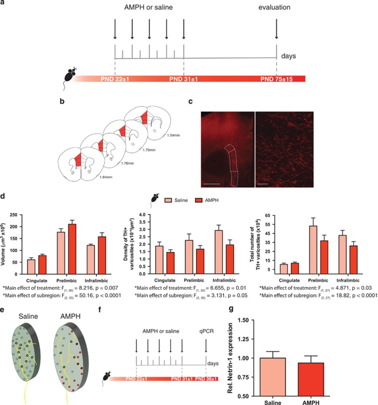
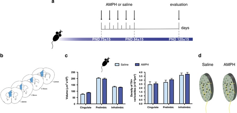
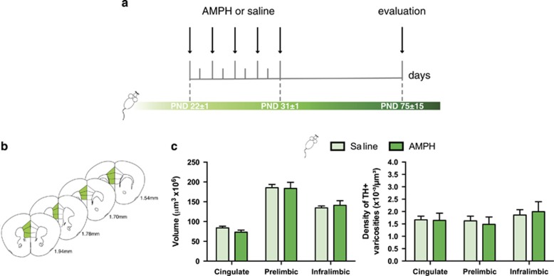
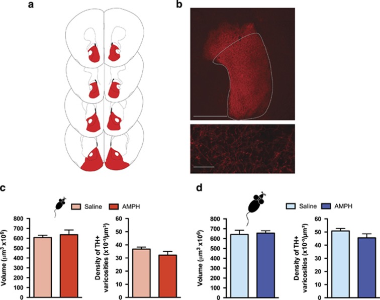
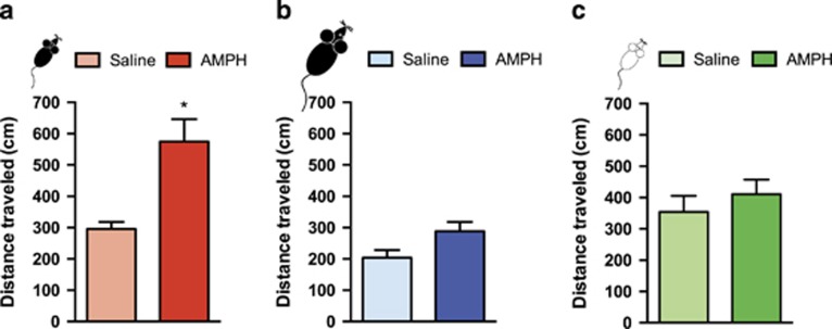
Initiation of drug use during adolescence is a strong predictor of both the incidence and severity of addiction throughout the lifetime. Intriguingly, adolescence is a period of dynamic refinement in the organization of neuronal connectivity, in particular medial prefrontal cortex (mPFC) dopamine circuitry. The guidance cue receptor, DCC (deleted in colorectal cancer), is highly expressed by dopamine neurons and orchestrates their innervation to the mPFC during adolescence. Furthermore, we have shown that amphetamine in adolescence regulates DCC expression in dopamine neurons. Drugs in adolescence may therefore induce their enduring behavioral effects via DCC-mediated disruption in mPFC dopamine development. In this study, we investigated the impact of repeated exposure to amphetamine during adolescence on both the development of mPFC dopamine connectivity and on salience attribution to drug context in adulthood. We compare these effects to those induced by adult exposure to an identical amphetamine regimen. Finally, we determine whether DCC signaling within dopamine neurons is necessary for these events. Exposure to amphetamine in adolescence, but not in adulthood, leads to an increase in the span of dopamine innervation to the mPFC, but a reduction of presynaptic sites present on these axons. Amphetamine treatment in adolescence, but not in adulthood, also produces an increase in salience attribution to a previously drug-paired context in adulthood. Remarkably, DCC signaling within dopamine neurons is required for both of these effects. Drugs of abuse in adolescence may therefore induce their detrimental behavioral consequences by disrupting mesocortical dopamine development through alterations in the DCC signaling cascade.
Figures
Similar articles
-
Resilience to amphetamine in mouse models of netrin-1 haploinsufficiency: role of mesocortical dopamine.Psychopharmacology (Berl). 2015 Oct;232(20):3719-29. doi: 10.1007/s00213-015-4032-9. Epub 2015 Aug 12. Psychopharmacology (Berl). 2015. PMID: 26264903
-
DCC-related developmental effects of abused- versus therapeutic-like amphetamine doses in adolescence.Addict Biol. 2020 Jul;25(4):e12791. doi: 10.1111/adb.12791. Epub 2019 Jun 13. Addict Biol. 2020. PMID: 31192517 Free PMC article.
-
Dcc haploinsufficiency regulates dopamine receptor expression across postnatal lifespan.Neuroscience. 2017 Mar 27;346:182-189. doi: 10.1016/j.neuroscience.2017.01.009. Epub 2017 Jan 17. Neuroscience. 2017. PMID: 28108253 Free PMC article.
-
Making Dopamine Connections in Adolescence.Trends Neurosci. 2017 Dec;40(12):709-719. doi: 10.1016/j.tins.2017.09.004. Epub 2017 Oct 9. Trends Neurosci. 2017. PMID: 29032842 Free PMC article. Review.
-
Role of netrin-1 in the organization and function of the mesocorticolimbic dopamine system.J Psychiatry Neurosci. 2011 Sep;36(5):296-310. doi: 10.1503/jpn.100171. J Psychiatry Neurosci. 2011. PMID: 21481303 Free PMC article. Review.
Cited by
-
AMPed-up adolescents: The role of age in the abuse of amphetamines and its consequences on cognition and prefrontal cortex development.Pharmacol Biochem Behav. 2020 Nov;198:173016. doi: 10.1016/j.pbb.2020.173016. Epub 2020 Aug 20. Pharmacol Biochem Behav. 2020. PMID: 32828971 Free PMC article. Review.
-
Resilience to amphetamine in mouse models of netrin-1 haploinsufficiency: role of mesocortical dopamine.Psychopharmacology (Berl). 2015 Oct;232(20):3719-29. doi: 10.1007/s00213-015-4032-9. Epub 2015 Aug 12. Psychopharmacology (Berl). 2015. PMID: 26264903
-
Amphetamines in child medicine: a review of ClinicalTrials.gov.Front Pharmacol. 2023 Oct 3;14:1280562. doi: 10.3389/fphar.2023.1280562. eCollection 2023. Front Pharmacol. 2023. PMID: 37854716 Free PMC article.
-
Low-cost conditioned place preference setup including video recording and analysis of behaviour.MethodsX. 2020 Apr 25;7:100899. doi: 10.1016/j.mex.2020.100899. eCollection 2020. MethodsX. 2020. PMID: 32405466 Free PMC article.
-
Δ-Tetrahydrocannabinol Increases Dopamine D1-D2 Receptor Heteromer and Elicits Phenotypic Reprogramming in Adult Primate Striatal Neurons.iScience. 2020 Jan 24;23(1):100794. doi: 10.1016/j.isci.2019.100794. Epub 2019 Dec 24. iScience. 2020. PMID: 31972514 Free PMC article.
References
-
- Anggård E, Gunne LM, Jönsson L-E, Niklasson F. Pharmacokinetic and clinical studies on amphetamine dependent subjects. Eur J Clin Pharmacol. 1970;3:3–11.
-
- Anthony JC, Petronis KR. Early-onset drug use and risk of later drug problems. Drug and Alcohol Depend. 1995;40:9–15. - PubMed
-
- Antonopoulos J, Dori I, Dinopoulos A, Chiotelli M, Parnavelas JG. Postnatal development of the dopaminergic system of the striatum in the rat. Neuroscience. 2002;110:245–256. - PubMed
-
- Benes FM, Taylor JB, Cunningham MC. Convergence and plasticity of monoaminergic systems in the medial prefrontal cortex during the postnatal period: implications for the development of psychopathology. Cereb Cortex. 2000;10:1014–1027. - PubMed
-
- Benes FM, Vincent SL, Molloy R, Khan Y. Increased interaction of dopamine-immunoreactive varicosities with GABA neurons of rat medial prefrontal cortex occurs during the postweanling period. Synapse. 1996;23:237–245. - PubMed
Publication types
MeSH terms
Substances
Grants and funding
LinkOut - more resources
Full Text Sources
Other Literature Sources
