

Cocoon-like self-degradable DNA nanoclew for anticancer drug delivery
- PMID: 25336272
- PMCID: PMC4210150
- DOI: 10.1021/ja5088024
Cocoon-like self-degradable DNA nanoclew for anticancer drug delivery
Abstract
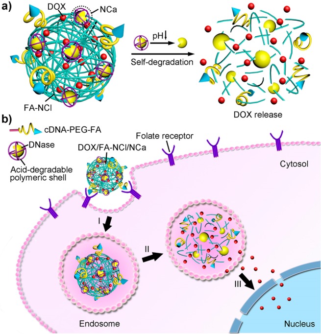
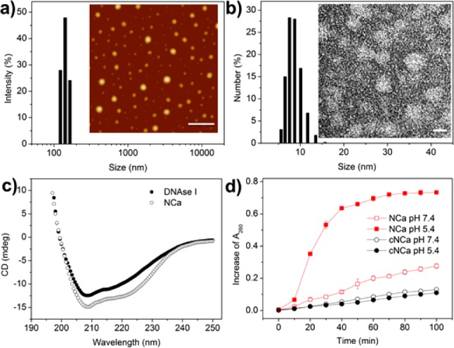
A bioinspired cocoon-like anticancer drug delivery system consisting of a deoxyribonuclease (DNase)-degradable DNA nanoclew (NCl) embedded with an acid-responsive DNase I nanocapsule (NCa) was developed for targeted cancer treatment. The NCl was assembled from a long-chain single-stranded DNA synthesized by rolling-circle amplification (RCA). Multiple GC-pair sequences were integrated into the NCl for enhanced loading capacity of the anticancer drug doxorubicin (DOX). Meanwhile, negatively charged DNase I was encapsulated in a positively charged acid-degradable polymeric nanogel to facilitate decoration of DNase I into the NCl by electrostatic interactions. In an acidic environment, the activity of DNase I was activated through the acid-triggered shedding of the polymeric shell of the NCa, resulting in the cocoon-like self-degradation of the NCl and promoting the release of DOX for enhanced therapeutic efficacy.
Figures


References
-
- Lee H.; Lytton-Jean A. K. R.; Chen Y.; Love K. T.; Park A. I.; Karagiannis E. D.; Sehgal A.; Querbes W.; Zurenko C. S.; Jayaraman M.; Peng C. G.; Charisse K.; Borodovsky A.; Manoharan M.; Donahoe J. S.; Truelove J.; Nahrendorf M.; Langer R.; Anderson D. G. Nat. Nanotechnol. 2012, 7, 389–393. - PMC - PubMed
- Douglas S. M.; Bachelet I.; Church G. M. Science 2012, 335, 831–834. - PubMed
- Andersen E. S.; Dong M.; Nielsen M. M.; Jahn K.; Subramani R.; Mamdouh W.; Golas M. M.; Sander B.; Stark H.; Oliveira C. L. P.; Pedersen J. S.; Birkedal V.; Besenbacher F.; Gothelf K. V.; Kjems J. Nature 2009, 459, 73–76. - PubMed
- Zhang Z.; Eckert M. A.; Ali M. M.; Liu L.; Kang D.-K.; Chang E.; Pone E. J.; Sender L. S.; Fruman D. A.; Zhao W. ChemBioChem 2014, 15, 1268–1273. - PubMed
- Lo P. K.; Karam P.; Aldaye F. A.; McLaughlin C. K.; Hamblin G. D.; Cosa G.; Sleiman H. F. Nat. Chem. 2010, 2, 319–328. - PubMed
- Zhang Z.; Ali M. M.; Eckert M. A.; Kang D.-K.; Chen Y. Y.; Sender L. S.; Fruman D. A.; Zhao W. Biomaterials 2013, 34, 9728–9735. - PubMed
- Zhu G.; Hu R.; Zhao Z.; Chen Z.; Zhang X.; Tan W. J. Am. Chem. Soc. 2013, 135, 16438–16445. - PMC - PubMed
- Hu R.; Zhang X.; Zhao Z.; Zhu G.; Chen T.; Fu T.; Tan W. Angew. Chem., Int. Ed. 2014, 53, 5821–5826. - PubMed
-
- Wang K.; You M.; Chen Y.; Han D.; Zhu Z.; Huang J.; Williams K.; Yang C. J.; Tan W. Angew. Chem., Int. Ed. 2011, 50, 6098–6101. - PubMed
Publication types
MeSH terms
Substances
Grants and funding
LinkOut - more resources
Full Text Sources
Other Literature Sources
Miscellaneous
