. 2015 Feb 18;4(3):460-8.
doi: 10.1002/adhm.201400421. Epub 2014 Oct 21.

Plasmonic nanodiamonds: targeted core-shell type nanoparticles for cancer cell thermoablation

Affiliations

Plasmonic nanodiamonds: targeted core-shell type nanoparticles for cancer cell thermoablation

Ivan Rehor et al. Adv Healthc Mater. .

Abstract

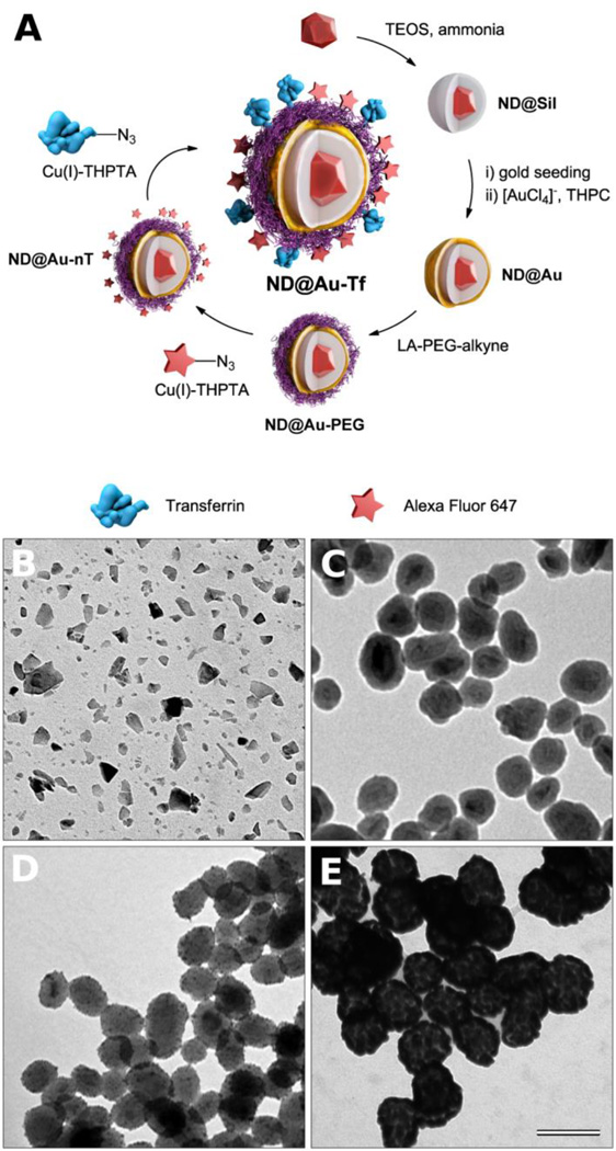
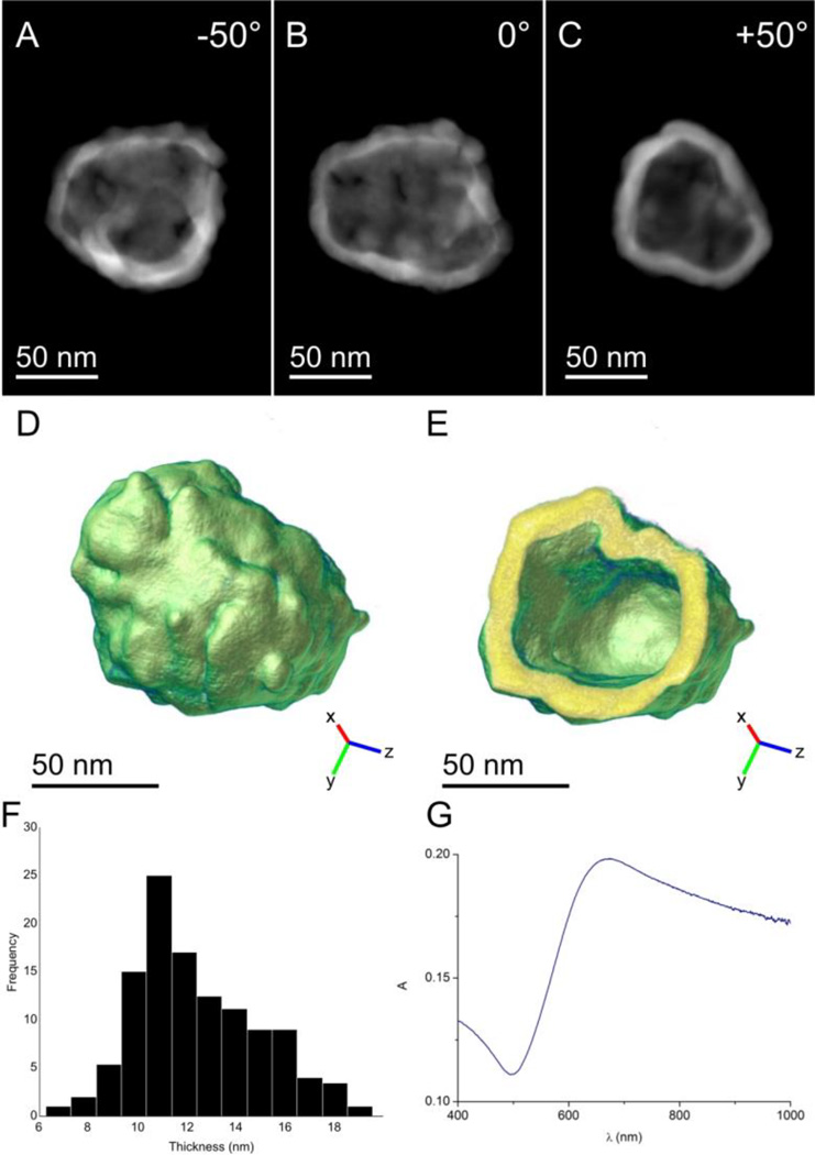
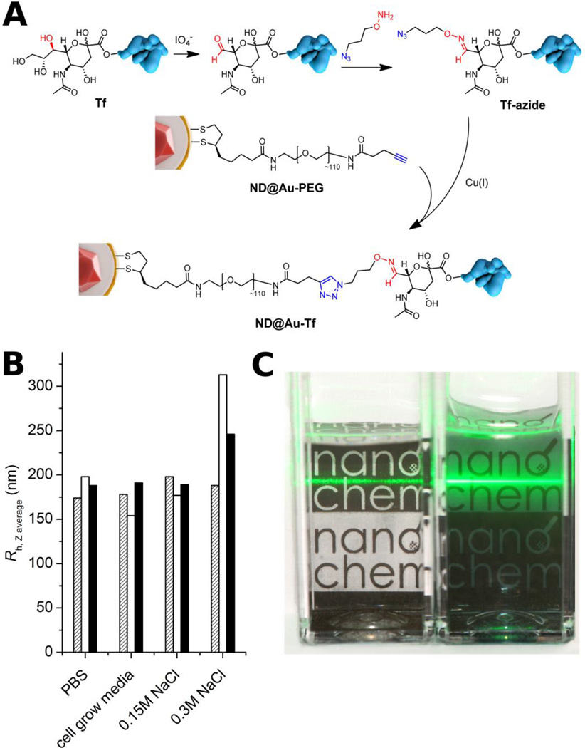
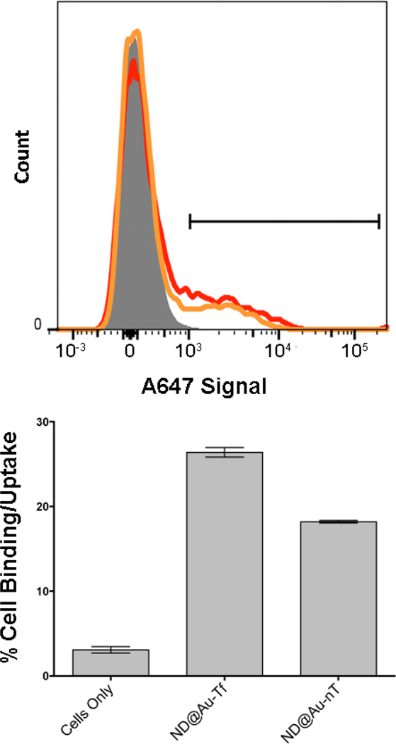
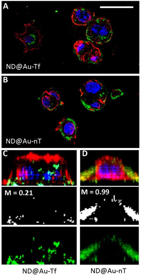
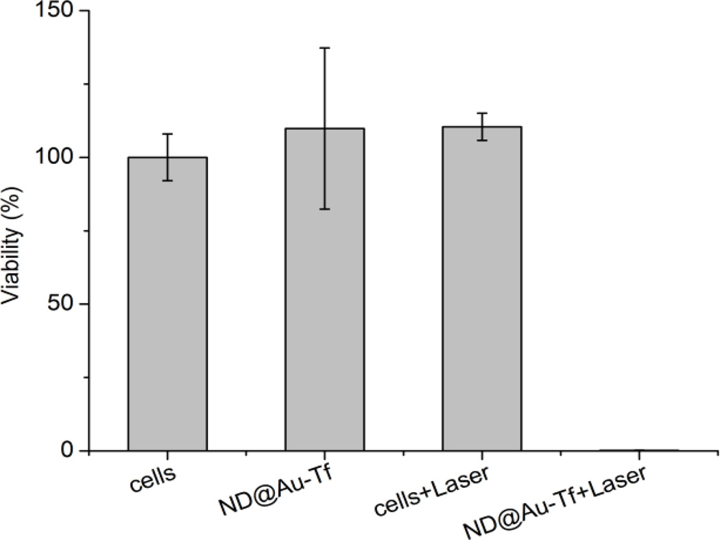
Targeted biocompatible nanostructures with controlled plasmonic and morphological parameters are promising materials for cancer treatment based on selective thermal ablation of cells. Here, core-shell plasmonic nanodiamonds consisting of a silica-encapsulated diamond nanocrystal coated in a gold shell are designed and synthesized. The architecture of particles is analyzed and confirmed in detail using electron tomography. The particles are biocompatibilized using a PEG polymer terminated with bioorthogonally reactive alkyne groups. Azide-modified transferrin is attached to these particles, and their high colloidal stability and successful targeting to cancer cells overexpressing the transferrin receptor are demonstrated. The particles are nontoxic to the cells and they are readily internalized upon binding to the transferrin receptor. The high plasmonic cross section of the particles in the near-infrared region is utilized to quantitatively ablate the cancer cells with a short, one-minute irradiation by a pulse 750-nm laser.

Keywords: ablation; cancer; gold; nanodiamonds; plasmonics.


Figures

Figure 1
Figure 1
(A) Schematic representation of the preparation of GNSs with a diamond core. First, a silica shell is created on diamond particles, followed by formation of a GNS upon reduction of [AuCl4] promoted by adsorbed gold nanoparticle seeds. The GNS is modified with a lipoic acid-PEG conjugate, which is terminated with an alkyne. Using click chemistry, Alexa Fluor 647 dye and azide-modified transferrin (the targeting protein) are attached in consecutive steps. (B–D) TEM microphotographs of (B) diamond particles, (C) silica-coated diamond particles (ND@Sil), (D) silica-coated diamond particles with gold seeds, and (E) GNSs a with diamond core (ND@Au). The magnification is the same for all microphotographs, and the scale bar corresponds to 100 nm.
Figure 2
Figure 2
(A–C) 2D HAADF-STEM projections of a ND-silica particle coated with a GNS (ND@Au) obtained at different tilt angles. The diamond core and silica coating are not visible due to the limited dynamic range of the image detector. (D) A 3D representation of the reconstructed nanoshell. (E) A slice through the 3D reconstruction of the GNS demonstrating the homogeneity of shell thickness. (F) A histogram indicating the measured thicknesses of the shell based on electron tomography reconstruction. The average shell thickness was 12.6 ± 0.3 nm. (G) Absorption spectrum of ND@Au in water at 15 µg/mL concentration (which corresponds to a ND concentration of 0.5 µg/mL).
Figure 3
Figure 3
Structure of ND@Au-PEG conjugate and its colloidal stability in aqueous solutions with high ionic strength. A) Composition of the particle surface architecture after modification and attachment of Tf. B) Hydrodynamic radii of ND@Au-PEG in various solutions after 1 h (hatched), 1 week (white) and 1 month (black), showing no aggregation. C) Photograph of naked (ND@Au, left) and PEG-coated (ND@Au-PEG, right) particles dispersed in PBS (20 min after mixing; 0.2 mg/mL concentration). The precipitating ND@Au particles are already partially sedimented on the bottom of the vial, while the remaining large aggregates unevenly scatter the laser beam. The ND@Au-PEG particles form a stable colloidal solution, which evenly and strongly scatters the laser beam.
Figure 4
Figure 4
SKBR3 cell interactions with particles determined by flow cytometry. Upper histogram: gray, cells only; red, ND@Au-Tf; orange, ND@Au-nT. Bottom graph: Statistical analysis showing percent cellular uptake (positive cells shown on histogram by gate) for each sample. Error bars indicate standard deviation. Experiments were conducted in triplicate, and 10,000 gated events were analyzed. Student’s t-test indicates significant differences comparing targeted ND@Au-Tf and non-targeted ND@Au-nT formulations (p < 0.05).
Figure 5
Figure 5
SKBR3 cell interactions with particles observed by confocal microscopy. The particles were incubated with SKBR3 cells for 16 h, fixed, stained, and imaged. ND@Au-Tf (A+C) and ND@Au-nT (B+D) are pseudo-colored in green (imaged based on the Alexa Fluor 647 label), nuclei are shown in blue (stained with DAPI), and cell membranes are shown in red (stained with WGA-A555); the scale bar is 30 µm. C+D shows 3D reconstruction of single cells: the top panel shows all channels, the middle panel depicts co-localization of the particles and WGA signals (M = Mander’s coefficient of co-localization determined using ImageJ software), and the bottom panel shows ND signals.
Figure 6
Figure 6
Laser ablation of HeLa cells incubated with ND@Au-Tf nanoparticles. Cell viability was estimated by luciferase assay with 24 h delay after 1 min irradiation with 37 W/cm2 intensity. The viability of cells treated with ND@Au-Tf and laser was ~0.15%.

References

    1. Khlebtsov NG, Dykman LA. J. Quant. Spectrosc. Radiat. Transf. 2010;111:1.
    1. Jones MR, Osberg KD, Macfarlane RJ, Langille MR, Mirkin CA. Chem. Rev. 2011;111:3736. - PubMed
    1. Daniel MC, Astruc D. Chem. Rev. 2004;104:293. - PubMed
    1. Henry A-I, Bingham JM, Ringe E, Marks LD, Schatz GC, Van Duyne RP. J. Phys. Chem. C. 2011;115:9291.
    1. Agrawal A, Huang S, Wei Haw Lin A, Barton JK, Drezek RA, Pfefer TJ, Lee M-H. J. Biomed. Opt. 2006;11:041121. - PubMed

Publication types

MeSH terms

LinkOut - more resources