. 2014 Dec;20(12):1878-89.
doi: 10.1261/rna.045633.114. Epub 2014 Oct 21.

Cellular microRNAs up-regulate transcription via interaction with promoter TATA-box motifs

Affiliations

Cellular microRNAs up-regulate transcription via interaction with promoter TATA-box motifs

Yijun Zhang et al. RNA. 2014 Dec.

Abstract

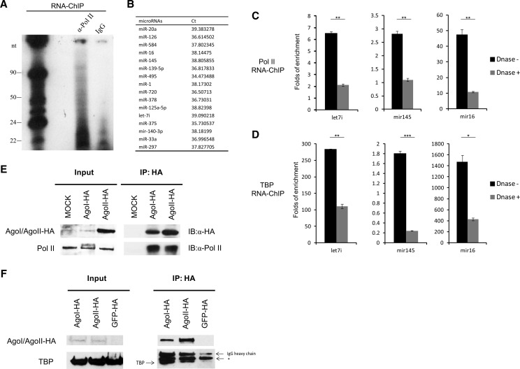
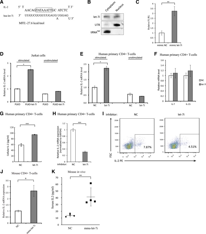
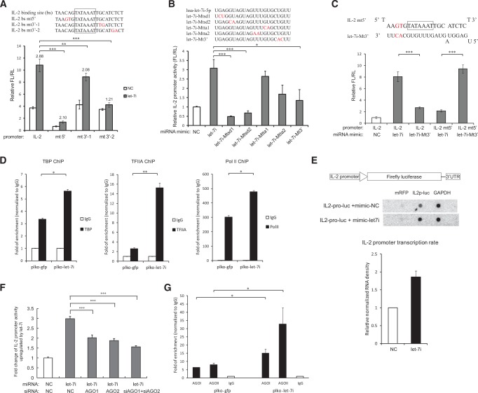
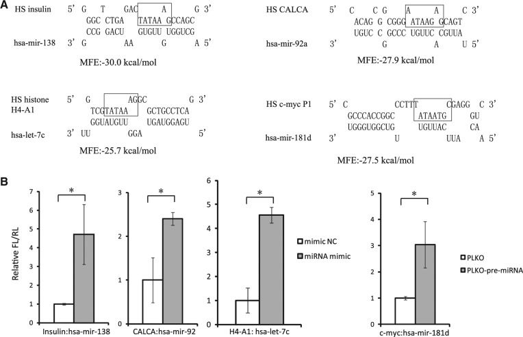
The TATA box represents one of the most prevalent core promoters where the pre-initiation complexes (PICs) for gene transcription are assembled. This assembly is crucial for transcription initiation and well regulated. Here we show that some cellular microRNAs (miRNAs) are associated with RNA polymerase II (Pol II) and TATA box-binding protein (TBP) in human peripheral blood mononuclear cells (PBMCs). Among them, let-7i sequence specifically binds to the TATA-box motif of interleukin-2 (IL-2) gene and elevates IL-2 mRNA and protein production in CD4(+) T-lymphocytes in vitro and in vivo. Through direct interaction with the TATA-box motif, let-7i facilitates the PIC assembly and transcription initiation of IL-2 promoter. Several other cellular miRNAs, such as mir-138, mir-92a or mir-181d, also enhance the promoter activities via binding to the TATA-box motifs of insulin, calcitonin or c-myc, respectively. In agreement with the finding that an HIV-1-encoded miRNA could enhance viral replication through targeting the viral promoter TATA-box motif, our data demonstrate that the interaction with core transcription machinery is a novel mechanism for miRNAs to regulate gene expression.

Keywords: TATA-box motif; core transcription machinery; microRNAs; transcription activation.


Figures

FIGURE 1.
FIGURE 1.
Cellular miRNAs and AGO proteins are associated with RNA Pol II core transcription machinery. (A) Autoradiogram of 32P-labeled RNA associated with human RNA Pol II purified by RNA chromatin immunoprecipitation (RNA-ChIP). The PBMCs were isolated from healthy individuals and activated with anti-CD3 (1 μg/mL) and anti-CD28 (5 μg/mL) antibodies for 48 h. RNA-ChIP assay was then performed. The chromatin-associated RNA was extracted and radiolabeled. These data represent three independent experiments. (B) Identification of RNA Pol II-associated miRNAs with miRNA expression microarray. The table only shows the detected miRNAs. qRT-PCR quantification of the miRNAs associated with RNA Pol II (C) and TBP (D) with or without DNase treatment. The enriched miRNAs were determined by real-time qRT-PCR and normalized to that of IgG. P-values were calculated using the two tailed unpaired Student's t-test with equal variances, n = 3 biological replicates. (*) P < 0.05, (**) P < 0.01, (***) P < 0.001. Co-immunoprecipitation (co-IP) assay to detect the association of HA-tagged Ago proteins with RNA Pol II (E) or TBP (F). HA-tagged Ago1 and Ago2 constructs were transfected into HEK293T cells. After 48 h, cell lysates were immunoprecipitated with anti-HA antibody, and subsequently detected with anti-RNA Pol II or anti-TBP antibody. (*) Nonspecific band. These data represent at least three independent experiments.
FIGURE 2.
FIGURE 2.
Let-7i enhances IL-2 promoter activity in human and mouse primary CD4+ T cells. (A) Predicted binding between let-7i and IL-2 core promoter region. TATA-box motif was highlighted. (B) Northern blot demonstrating the relative nuclear and cytoplasmic abundance of endogenous let-7i in Sup-T1 cells. U78 small nucleolar RNA and valine-tRNA (tRNAval) served as controls for the nuclear and cytoplasmic fractionation. (C) The let-7i mimic or negative control was co-transfected with the IL-2 promoter-driven firefly luciferase (FL) construct into HEK293T cells, a CMV-renilla luciferase (RL) plasmid served as a transfection control. Dual-luciferase assay was performed at 24 h after transfection. (D) The effect of overexpressing let-7i on IL-2 mRNA expression. The PLKO-let-7i cells which expressed let-7i miRNA plus GFP and PLKO cells which just expressed GFP were generated by infecting Jurkat cells with lentiviral vector containing let-7i precursor or the PLKO empty vector and sorted with FACS. The gfp-positive cells were subsequently treated with (left) and without (right) phorbolmyristate acetate (PMA, 50 ng/mL) and ionomycin (1 μM) for 12 h. IL-2 mRNA was then evaluated by qRT-PCR and normalized to GAPDH mRNA. (E) Levels of IL-2 mRNA in primary human CD4+ T cells after treatment with agomir-let-7i. The CD4+ T-cells were treated with agomir-let-7i or control for 48 h, and then cells were activated with anti-CD3 and anti-CD28 antibodies. The levels of IL-2 mRNA were evaluated by qRT-PCR and normalized to β-actin mRNA. (F) Levels of IL-7 and IL-15 mRNA in primary human CD4+ T cells after the treatment of agomir-let-7i as described above. (G) Cellular IL-2 protein was determined by ELISA. The samples were collected from the cell lysates of primary human CD4+ T cells transfected with let-7i mimic or negative control and activated as described above. (H) IL-2 mRNA levels in primary human CD4+ T cells transfected with let-7i miRNA inhibitor or negative control. The CD4+ T cells were transfected with let-7i inhibitor or negative control at a final concentration of 200 nM and then activated as described above. The levels of IL-2 mRNA were evaluated by qRT-PCR and normalized to GAPDH mRNA. (I) Intracellular staining of IL-2 in the CD+ cells transfected with let-7i miRNA inhibitor or negative control. The cells were transfected with let-7i miRNA inhibitor or negative control at 200 nM and activated with anti-CD3/anti-CD28 for 48 h before IL-2 intracellular staining and flow cytometry assay. These data represent three independent experiments. (J) IL-2 mRNA levels in mouse CD4+ T cells transfected with mmu-let-7i mimic or negative control. (K) Quantification of serum IL-2 levels of mice treated with agomir-mmu-let-7i or negative control (25 nmol per animal) for 48 h by ELISA. P-values were calculated using the two-tailed unpaired Student's t-test with equal variances, n = 3 biological replicates unless specified. (*) P < 0.05, (**) P < 0.01, (***) P < 0.001.
FIGURE 3.
FIGURE 3.
Sequence-specific binding between let-7i miRNA and the TATA-box motif facilitates IL-2 PIC assembly and transcription initiation. (A, top) Mutations were introduced into the predicted let-7i–binding site in IL-2 core promoter region (in red). (Bottom) Wild-type and mutated IL-2 promoter–driven reporter constructs were transfected into HEK293T cells with let-7i mimic or negative control. Cells were harvested at 36 h post transfection and the promoter activities were determined by dual-luciferase assay. Up-regulation folds of the promoter activities relative to the control by let-7i were shown. n = 5 biological replicates. (B) A series of mutations were generated in the 5′ seed region, the sequence complementary to IL-2 TATA box, or 3′ region of let-7i miRNA, respectively (mutated nucleotide in red). The wild-type let-7i mimic or these mutated mimics were transfected to HEK293T cells, respectively, with the IL-2 promoter–driven luciferase construct. The IL-2 promoter activities were examined as described above. n = 5 biological replicates. (C) A mutated let-7i mimic was co-transfected with an IL-2 promoter–driven reporter which harbored mutations base-pairing complementary to the mutated let-7i mimic, and promoter activities were measured as described above. n = 5 biological replicates. (D) ChIP assay with antibody against TBP, TFIIA, or Pol II in Jurkat cells expressing GFP only (plko-gfp) or let-7i precursor (plko-let-7i) to examine the IL-2 promoter DNA associated with the general transcription factors. The enrichment of the promoter DNA by the transcription factors was normalized to IgG. These data represent three independent experiments. (E) Nuclear run-on assay of IL-2 promoter activity. The IL-2 promoter–driven luciferase construct was co-transfected into HEK293T cells with let-7i precursor or the empty vector. GAPDH was selected as an input control and mRFP as negative control. This figure represents three independent experiments. (F) Effects of inhibiting Ago1, Ago2, or both genes on the activation of IL-2 promoter by let-7i miRNA mimics. Ago proteins were knockdown with specific siRNAs in HEK293T cells, and the promoter activities of IL-2 in the presence of let-7i or control mimic were measured as described above. n = 3 biological replicates. (G) ChIP experiments were performed with antibody against AGO1 or AGO2 in Jurkat cells expressing GFP (plko-gfp) or let-7i precursor (plko-let-7i) to examine their association with the IL-2 promoter. The plko-gfp cells and plko-let-7i cells were generated as described above. The primers covering -146 to -8 relative to the TSS of IL-2 promoter were used to detect the associated promoter DNA. The enrichments of the promoter DNA by Ago proteins were normalized to IgG. This figure represents three independent experiments. P-values were calculated using the two-tailed unpaired Student's t-test with equal variances. (*) P < 0.05, (**) P < 0.01, (***) P < 0.001.
FIGURE 4.
FIGURE 4.
TATA-box motif-specific miRNAs enhance the promoter activity of many important cellular genes. (A) Predicted interactions between the core promoter of four human genes and four human miRNAs. TATA-box motifs were highlighted. (B) The promoter activities of indicated genes were measured with dual-luciferase assay upon the overexpression of their corresponding miRNAs. The promoter-driven luciferase reporters were transfected into HEK293T cells with synthesized miRNA mimics or miRNA precursors containing construct. P-values were calculated using the two-tailed unpaired Student's t-test with equal variances, n = 3 biological replicates. (*) P < 0.05.

References

    1. Bartel DP 2004. MicroRNAs: genomics, biogenesis, mechanism, and function. Cell 116: 281–297. - PubMed
    1. Bartel DP 2009. MicroRNAs: target recognition and regulatory functions. Cell 136: 215–233. - PMC - PubMed
    1. Burley SK, Roeder RG 1996. Biochemistry and structural biology of transcription factor IID (TFIID). Annu Rev Biochem 65: 769–799. - PubMed
    1. Bushati N, Cohen SM 2007. microRNA functions. Annu Rev Cell Dev Biol 23: 175–205. - PubMed
    1. Cernilogar FM, Onorati MC, Kothe GO, Burroughs AM, Parsi KM, Breiling A, Lo Sardo F, Saxena A, Miyoshi K, Siomi H, et al.2011. Chromatin-associated RNA interference components contribute to transcriptional regulation in Drosophila. Nature 480: 391–395. - PMC - PubMed

Publication types

MeSH terms

LinkOut - more resources