. 2014 Dec 5;289(49):34229-40.
doi: 10.1074/jbc.M114.607366. Epub 2014 Oct 21.

Functionally important carboxyls in a bacterial homologue of the vesicular monoamine transporter (VMAT)

Affiliations

Functionally important carboxyls in a bacterial homologue of the vesicular monoamine transporter (VMAT)

Dana Yaffe et al. J Biol Chem. .

Abstract

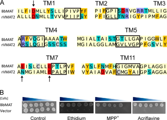
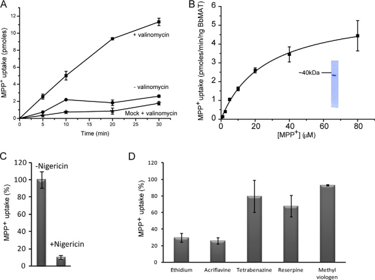
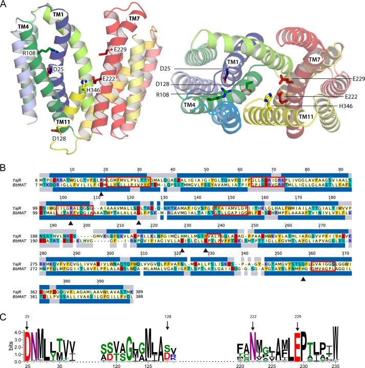
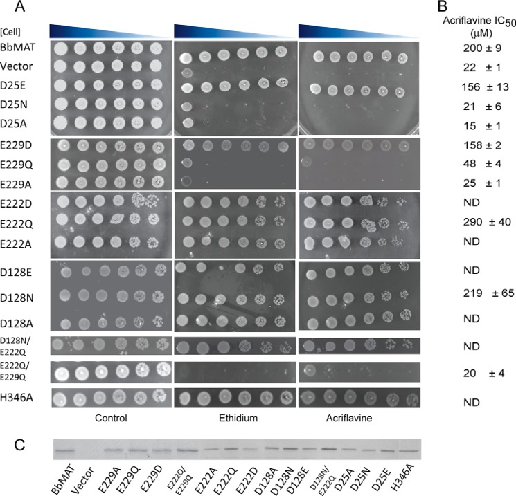
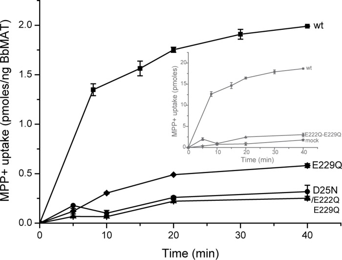
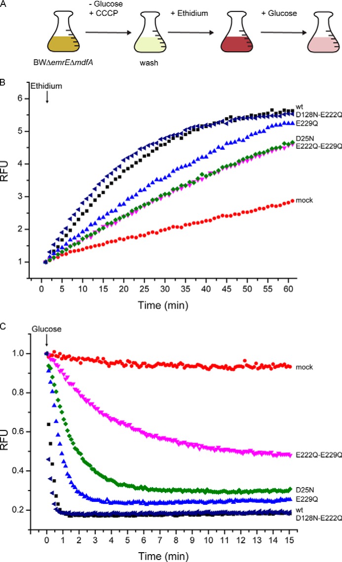
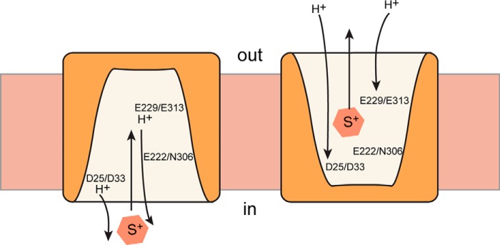
Transporters essential for neurotransmission in mammalian organisms and bacterial multidrug transporters involved in antibiotic resistance are evolutionarily related. To understand in more detail the evolutionary aspects of the transformation of a bacterial multidrug transporter to a mammalian neurotransporter and to learn about mechanisms in a milieu amenable for structural and biochemical studies, we identified, cloned, and partially characterized bacterial homologues of the rat vesicular monoamine transporter (rVMAT2). We performed preliminary biochemical characterization of one of them, Brevibacillus brevis monoamine transporter (BbMAT), from the bacterium B. brevis. BbMAT shares substrates with rVMAT2 and transports them in exchange with >1H(+), like the mammalian transporter. Here we present a homology model of BbMAT that has the standard major facilitator superfamily fold; that is, with two domains of six transmembrane helices each, related by 2-fold pseudosymmetry whose axis runs normal to the membrane and between the two halves. The model predicts that four carboxyl residues, a histidine, and an arginine are located in the transmembrane segments. We show here that two of the carboxyls are conserved, equivalent to the corresponding ones in rVMAT2, and are essential for H(+)-coupled transport. We conclude that BbMAT provides an excellent experimental paradigm for the study of its mammalian counterparts and bacterial multidrug transporters.

Keywords: Antibiotic Resistance; Membrane Transport; Multidrug Transporter; Neurotransmitter; Neurotransmitter Transport; Proton Transport.


Figures

FIGURE 1.
FIGURE 1.
BbMAT is a multidrug resistance transporter homologous to rVMAT2. A, sequence alignment of key regions demonstrating the conservation between rVMAT2 and BbMAT. The alignment is colored according to the chemical properties of the residues: aliphatic (Ala, Gly, Ile, Leu, Met, Pro, and Val) in pale yellow, polar uncharged (Asn, Gln, Ser, and Thr) in teal, aromatic (Phe, Trp, and Tyr) in yellow/orange, acidic (Asp and Glu) in red, basic (Lys and Arg) in blue, His in pale green, and Cys in gray. Red rectangles mark the MFS and DHA12 motifs (10, 25). Arrows indicate residues studied in this work. B, BbMAT confers resistance against ethidium, MPP+, and acriflavine. E. coli JM109 cells harboring BbMAT or empty vector were assayed for drug resistance. 4.5 μl of serial 10-fold dilutions of cells were spotted on plates supplemented with ethidium (100 μg/ml), MPP+ (3 mm), or acriflavine (100 μg/ml). The plates are representative of at least three independent experiments.
FIGURE 2.
FIGURE 2.
BbMAT catalyzes exchange of cationic substrates with protons. Proteoliposomes (1.5 μl) reconstituted with 9 ng of purified BbMAT were assayed for [3H]MPP+ uptake. A, transport time course with (■) and without (●) the K+ ionophore valinomycin. Also shown for reference (★) are liposomes reconstituted with no protein (mock). B, rates of transport in the presence of increasing concentrations of MPP+. The calculated Km value is 25 ± 2 μm, and Vmax is 5 ± 0.1 pmol/min/ng BbMAT protein. Inset, purified BbMAT analyzed by SDS-PAGE and visualized with Coomassie. C, inhibition of MPP+ transport by nigericin. D, inhibition of MPP+ transport by 10 μm of known substrates (ethidium, acriflavine, methyl viologen) or inhibitors (tetrabenazine, reserpine) of multidrug resistance transporters. Results presented are from duplicate experiments. Error bars indicate mean ± S.E. Experiments were repeated at least twice.
FIGURE 3.
FIGURE 3.
Structural model of BbMAT on the basis of the YajR transporter structure. A, the BbMAT model in an outward-facing conformation is represented as helical cartoons, viewed from the plane of the membrane with the periplasm at the top (left, with TM4 and part of TM11 omitted for clarity) and from the periplasm (right). The model predicts four negatively charged and two positively charged residues exposed to the central cavity, which could be involved in the transport activity of BbMAT. Side chains of these residues are shown as sticks. The figures were generated using PyMOL v1.7.05 (Schrödinger). B, sequence alignment between YajR and BbMAT used for modeling. The alignment was colored according to the chemical nature of the residues as described in Fig. 1. The helices assigned for the YajR structure using DSSP (67) and the PSIPRED prediction for helices in BbMAT are shown as blue bars above and below the sequences, respectively. Red rectangles mark the MFS and DHA12 motifs (10, 25), and black triangles indicate the residues that have been investigated in this study. C, sequence logo illustrating conservation of Asp-25, Asp-128, Glu-222, and Glu-229 among 500 homologues belonging to the MFS family. Multiple sequence alignment was performed using Muscle, and the logo was generated using Weblogo 3.3 (68). The residue colors are as follows: polar (Gly, Ser, Thr, Tyr, and Cys) in green, neutral (Gln and Asn) in purple, basic (Lys, Arg, and His) in blue, acidic (Asp and Glu) in red, and hydrophobic (Ala, Val, Leu, Ile, Pro, Trp, Phe, and Met) in black. Numbering is according to the sequence of BbMAT.
FIGURE 4.
FIGURE 4.
Two carboxyl groups are important for conferring resistance by BbMAT. A, drug resistance of E. coli cells harboring the indicated BbMAT mutant. Serial dilutions of cells were spotted on plates supplemented with ethidium (100 μg/ml) or acriflavine (100 μg/ml). The plates are representative of at least three independent experiments. B, IC50 values for acriflavine were assessed by calculating the amount of drug required to inhibit cell growth by 50%, as described under “Experimental Procedures.” The experiments were repeated at least twice. C, expression of the different mutants assessed by Coomassie staining. ND, not determined.
FIGURE 5.
FIGURE 5.
[3H]MPP+ uptake into proteoliposomes. [3H]MPP+ uptake was assayed as described in the legend for Fig. 2, with liposomes prepared from wild-type (■) and D25N-expressing (♦), E229Q-expressing (●), and E222Q/E229Q-expressing (▴) cells. Inset, for comparison, the results not corrected for protein content are shown for the wild type (■), E222Q/E229Q (▴), and mock liposomes (★).
FIGURE 6.
FIGURE 6.
Asp-25 and Glu-229 are important for proton and substrate binding. A, schematic illustrating the protocol of the experiment. CCCP, carbonyl cyanide m-chlorophenylhydrazone. B and C, ethidium transport activity of BbMAT mutants measured using ethidium fluorescence and shown in relative fluorescence units (RFU). E. coli BWΔemrEΔmdfA cells expressing the wild type (black ■), D128N/E222Q (dark blue ◀), E229Q (light blue ▴), E222Q/E229Q (pink ▾), and D25N (green ▴) BbMAT or mock-transformed (red ●) were assayed for downhill passive ethidium uptake (B) and uphill efflux initiated by the addition of glucose (C). In C, the initial value of fluorescence for each strain was taken as 1.
FIGURE 7.
FIGURE 7.
A simplified view of the catalytic cycle of BbMAT and rVMAT2. For the sake of simplicity, the schematic includes only the conformations where the protein faces the inside (in) or the outside (out), with two protons moving to the inside of the cell in the case of BbMAT or to the vesicular lumen in the case of rVMAT2. In the schematic, the conserved amino acids in BbMAT and the corresponding ones in rVM AT2 are shown, and their hypothetical role in substrate and proton binding is presented.

References

    1. Eiden L. E. (2000) The vesicular neurotransmitter transporters: current perspectives and future prospects. FASEB J. 14, 2396–2400 - PubMed
    1. Schuldiner S., Shirvan A., Linial M. (1995) Vesicular neurotransmitter transporters: from bacteria to humans. Physiol. Rev. 75, 369–392 - PubMed
    1. Chaudhry F. A., Edwards R. H., Fonnum F. (2008) Vesicular neurotransmitter transporters as targets for endogenous and exogenous toxic substances. Annu. Rev. Pharmacol. Toxicol. 48, 277–301 - PubMed
    1. Edwards R. (1992) The transport of neurotransmitters into synaptic vesicles. Curr. Opin. Neurobiol. 2, 586–594 - PubMed
    1. Yelin R., Schuldiner S. (1995) The pharmacological profile of the vesicular monoamine transporter resembles that of multidrug transporters. FEBS Lett. 377, 201–207 - PubMed

Publication types

MeSH terms

Associated data