. 2015 Jan;66(1):283-91.
doi: 10.1093/jxb/eru418. Epub 2014 Oct 21.

MONOPTEROS directly activates the auxin-inducible promoter of the Dof5.8 transcription factor gene in Arabidopsis thaliana leaf provascular cells

Affiliations

MONOPTEROS directly activates the auxin-inducible promoter of the Dof5.8 transcription factor gene in Arabidopsis thaliana leaf provascular cells

Mineko Konishi et al. J Exp Bot. 2015 Jan.

Abstract

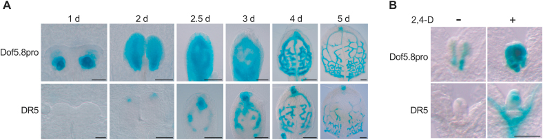
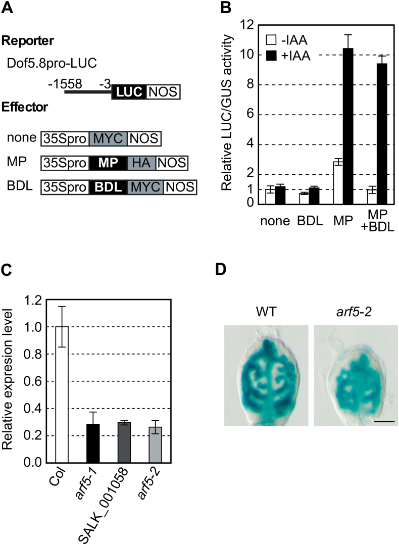
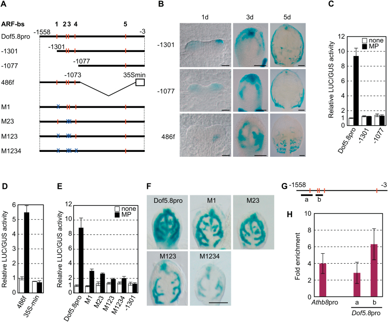
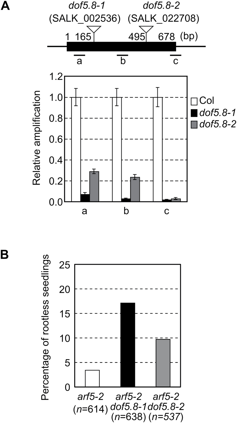
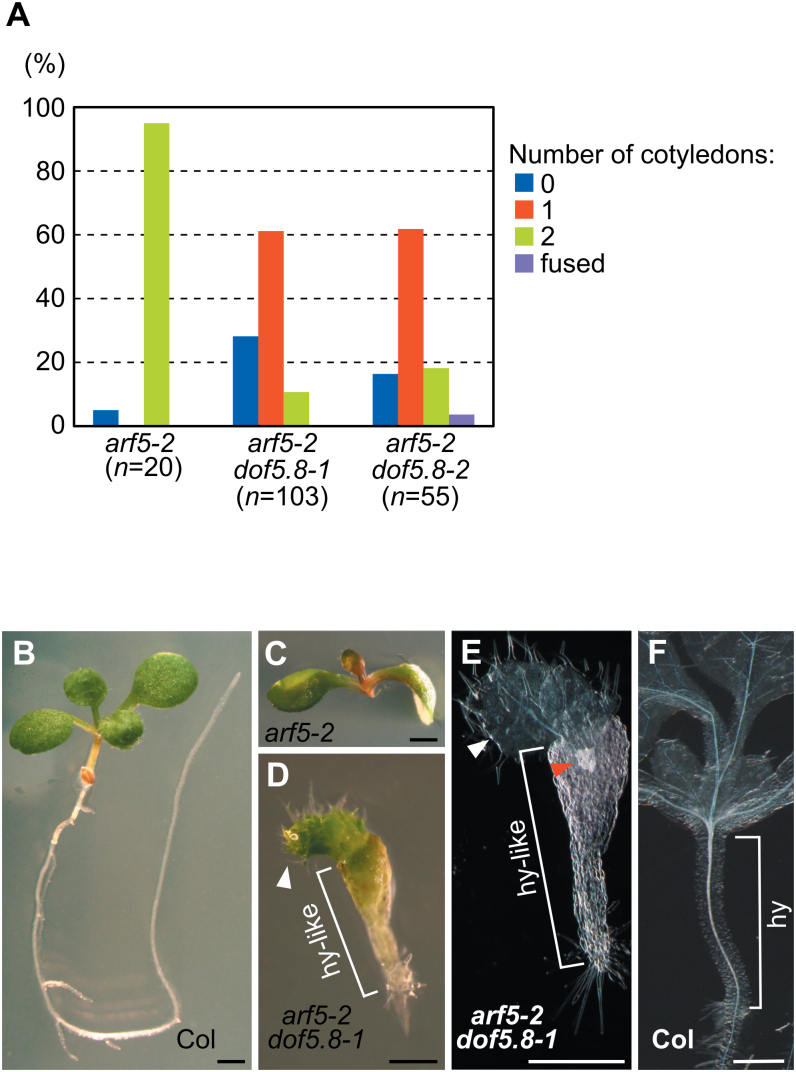
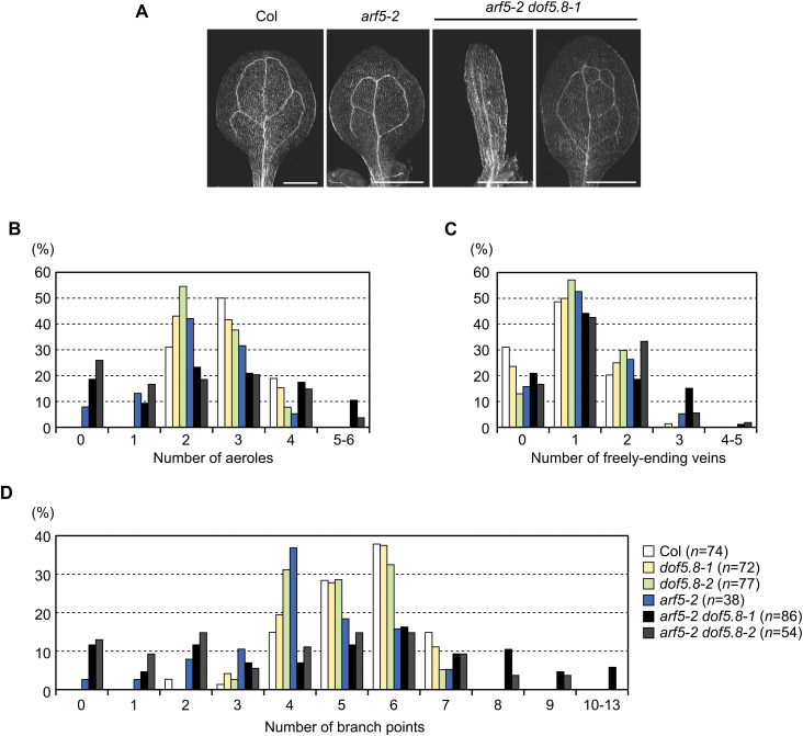
MONOPTEROS (MP) is an auxin-responsive transcription factor that is required for primary root formation and vascular development, whereas Dof5.8 is a Dof-class transcription factor whose gene is expressed in embryos as well as the pre- and procambial cells in the leaf primordium in Arabidopsis thaliana. In this study, it is shown that MP directly activates the Dof5.8 promoter. Although no apparent phenotype of the single dof5.8 mutants was found, phenotypic analysis with the mp dof5.8 double mutants revealed that mutations within Dof5.8 enhanced the phenotype of a weak allele of mp, with an increase in the penetrance of the 'rootless' phenotype and a reduction in the number of cotyledons. Furthermore, interestingly, although mp mutants showed reduced vascular pattern complexity in cotyledons, the mp dof5.8 double mutants displayed both more simplex and more complex vascular patterns in individual cotyledons. These results imply that the product of Dof5.8 whose expression is regulated by MP at least in part might be involved in multiple processes controlled by MP.

Keywords: Arabidopsis thaliana; Dof transcription factor; MONOPTEROS; auxin response; embryo development; vascular development..


Figures

Fig. 1.
Fig. 1.
Provascular activity of the Dof5.8 promoter and its enhancement by auxin. (A) Time course analysis of GUS expression under the control of the Dof5.8 promoter (Dof5.8pro) or the DR5 element (DR5) during development of the primordia of the first true leaves. Two primordia flanking the shoot apical meristem (1 d and 2 d) and individual leaf primordia (2.5–5 d) are shown. Scale bars=20 μm in 1 d and 2 d, 50 μm in 2.5 d and 3 d, 100 μm in 4 d and 5 d. (B) Effect of auxin treatment on the activity of the Dof5.8 promoter. Seedlings were treated or not with 10 μM 2,4-D for 16h. Scale bar=100 μm.
Fig. 2.
Fig. 2.
Activation of the Dof5.8 promoter by MP. (A) Schematic diagrams of the reporter and effector constructs used in the protoplast transactivation assay described in (B). Numbers indicate nucleotide positions relative to the translation start codon. ‘MYC’ and ‘HA’ are MYC- and haemagglutinin-tag peptides. (B) Effects of MP and auxin on the activity of the Dof5.8 promoter in protoplasts. The Dof5.8pro-LUC reporter construct was co-transfected with the expression plasmid of BDL, MP, or both, or an empty vector (none) and then protoplasts were incubated in the presence (+ IAA) or absence (– IAA) of 1 μM IAA. An internal control plasmid (UBQ10-GUS) was also co-transfected to normalize LUC reporter activity levels. Relative levels of LUC activity are shown as the means ±SD (n=3). (C) Relative Dof5.8 transcript levels in three mp alleles. Total RNA was extracted from the shoots of 4-day-old seedlings and used for qRT–PCR analysis. The transcript levels in wild-type A. thaliana (Col) were set to 1, as all three mp alleles (arf5-1, SALK_001058, and arf5-2) were in the Colombia background. Data are shown as the means ±SD (n=3). (D) GUS staining of the first true leaves of 4-day-old wild-type and arf5-2 seedlings that harbour the GUS gene under the control of the Dof5.8 promoter. The arf5-2 homozygous seedlings obtained from a segregating population of the plant homozygous for the Dof5.8pro-GUS transgene and heterozygous for the arf5-2 allele were used. Scale bar=50 μm.
Fig. 3.
Fig. 3.
Identification of MP-binding sites and the promoter region required for Dof5.8 expression in provascular cells. (A) Schematic representation of the promoter fragments used for deletion and mutational analyses of the Dof5.8 promoter. The sequences matching the ARF-binding consensus sequence (ARF-bs), and disruptions in these sequences are indicated by red bars and blue ‘X’s, respectively. Numbers indicate nucleotide positions relative to the translation start codons. A 486bp fragment from –1558 to –1073 was fused to the 35S minimal promoter truncated at –72 (35S min) to generate a fusion promoter (the 486f promoter). The mutated sequences are 5’-ACAGAG-3’ in ARF-bs 1–3 and 5’-ACAGTG-3’ in ARF-bs 4. (B) The activity of the truncated Dof5.8 promoters and the 486f promoter in the primordia of the first leaves. GUS staining of the first leaves of the transgenic seedlings carrying the GUS gene under the control of the deletion promoters described in (A). Scale bars=20 μm (1 d), 50 μm (3 d), and 100 μm (5 d). (C–E) MP-mediated transactivation of truncated Dof5.8 promoters (C), the 486f synthetic promoter (D), and mutated Dof5.8 promoters (E) in protoplasts. Promoters fused to LUC were co-transfected with the 35S-MP-HA plasmid (black bars) or an empty vector (white bars). Data are shown as the means ±SD (n=3). (F) GUS staining of the first leaves of the 4-day-old transgenic seedlings carrying the GUS gene under the control of the mutated promoters described in (A). Scale bar=100 μm. (G) Schematic representation of the Dof5.8 promoter showing ARF-binding sites (red bars) and the positions of the amplified DNA fragments (bars a and b) used in the ChIP-qPCR analysis shown in (H). (H) ChIP analysis of the binding of MP to the Dof5.8 promoter. Four-day-old transgenic seedlings expressing CFP-tagged MP were used. A DNA fragment from the Athb8 promoter was amplified as a positive control (Donner et al., 2009). The values were normalized using amplified DNA from a promoter unrelated to MP (the UBQ10 promoter).
Fig. 4.
Fig. 4.
Enhancement of the rootless phenotype of the arf5-2 mutant by dof5.8 mutations. (A) The positions of T-DNA insertions and transcript levels of Dof5.8 in the dof5.8-1 and dof5.8-2 mutant lines. The black box indicates the exon, and the positions of the three amplicons (a–c) are shown below. Nucleotide numbers are given relative to the translation start codon. The transcript levels in wild-type A. thaliana (Col) were set to 1. Data are shown as the means ±SD (n=3). (B) The percentages of the seedlings with the rootless phenotype from segregating populations of arf5-2 single and arf5-2 dof5.8 double mutants. Populations derived from parental plants heterozygous for the arf5-2 allele in the wild-type, dof5.8-1 homozygous or dof5.8-2 homozygous background were analysed.
Fig. 5.
Fig. 5.
The synergistic effects caused by dof5.8 and arf5-2 mutations. (A) The number of cotyledons in rootless seedlings of arf5-2 and arf5-2 dof5.8 mutants. (B–D) Images of 7-day-old wild-type (B), arf5-2 (C), and arf5-2 dof5.81 (D) seedlings. (E, F) Cleared images of arf5-2 dof5.8-1 (E) and Col (F) seedlings. White and red arrowheads in (D, E) indicate true leaves and vascular elements, respectively. ‘hy-like’ and ‘hy’ in (D–F) indicate a hypocotyl-like structure and hypocotyl, respectively. Scale bars=1mm in (B–D) and 0.5mm in (D–F).
Fig. 6.
Fig. 6.
The vascular patterns of arf5-2 dof5.8 cotyledons. (A) Representative images of the vascular pattern of cotyledons of wild-type (Col), arf5-2 mutant, and arf5-2 dof5.8-1 double mutant seedlings. Scale bars=1mm. (B–D) The percentage of cotyledons with the indicated number of aeroles (B), freely ending veins (C), and branch points (D). Seeds from plants heterozygous for arf5-2 in the wild-type, dof5.8-1 homozygous or dof5.8-2 homozygous background were sown, and rootless seedlings were used in this analysis. (This figure is available in colour at JXB online.)

References

    1. Alonso JM, Stepanova AN, Leisse TJ, et al. 2003. Genome-wide insertional mutagenesis of Arabidopsis thaliana . Science 301, 653–657. - PubMed
    1. Bauby H, Divol F, Truernit E, Grandjean O, Palauqui JC. 2007. Protophloem differentiation in early Arabidopsis thaliana development. Plant and Cell Physiology 48, 97–109. - PubMed
    1. Berleth T, Jürgens G. 1993. The role of the monopteros gene in organising the basal body region of the Arabidopsis embryo. Development 118, 575–587.
    1. Beuchat J, Scacchi E, Tarkowska D, Ragni L, Strnad M, Hardtke CS. 2010. BRX promotes Arabidopsis shoot growth. New Phytologist 188, 23–29. - PubMed
    1. Ckurshumova W, Scarpella E, Goldstein RS, Berleth T. 2011. Double-filter identification of vascular-expressed genes using Arabidopsis plants with vascular hypertrophy and hypotrophy. Plant Science 181, 96–104. - PubMed

Publication types

MeSH terms