. 2014 Nov 4;111(44):15756-61.
doi: 10.1073/pnas.1417898111. Epub 2014 Oct 21.

G protein-coupled receptor 56 regulates mechanical overload-induced muscle hypertrophy

Affiliations

G protein-coupled receptor 56 regulates mechanical overload-induced muscle hypertrophy

James P White et al. Proc Natl Acad Sci U S A. .

Abstract

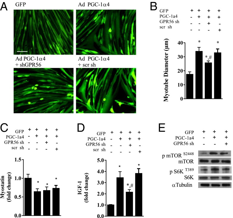
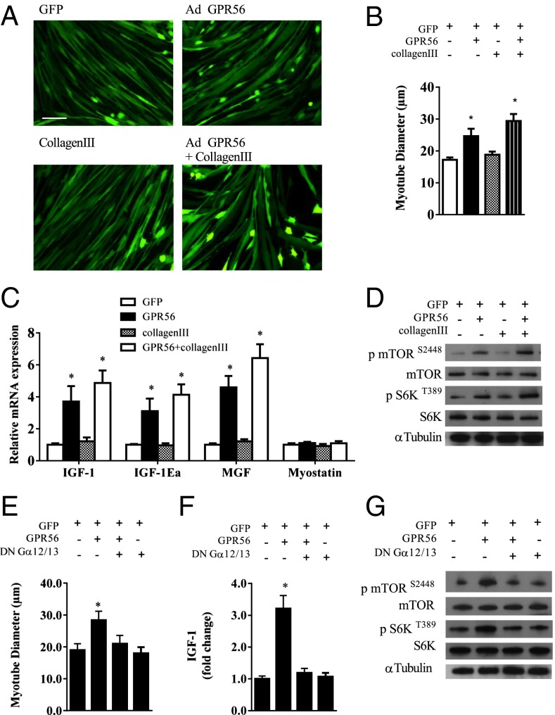
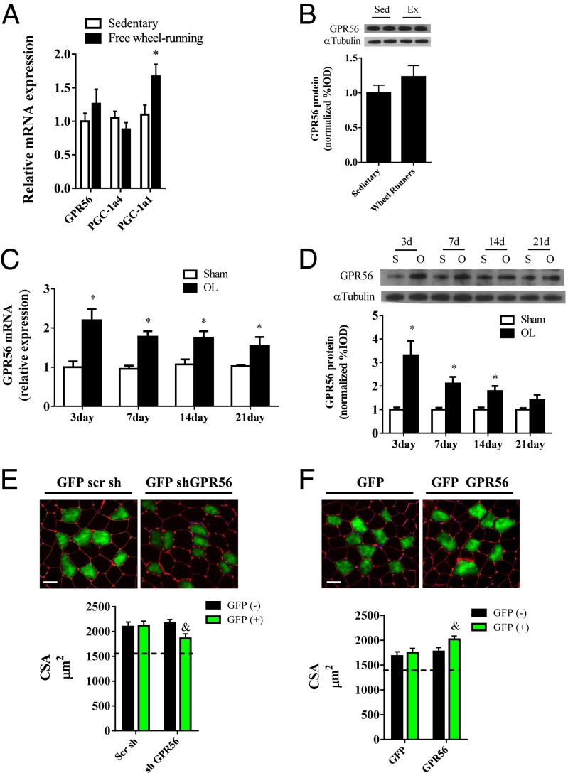
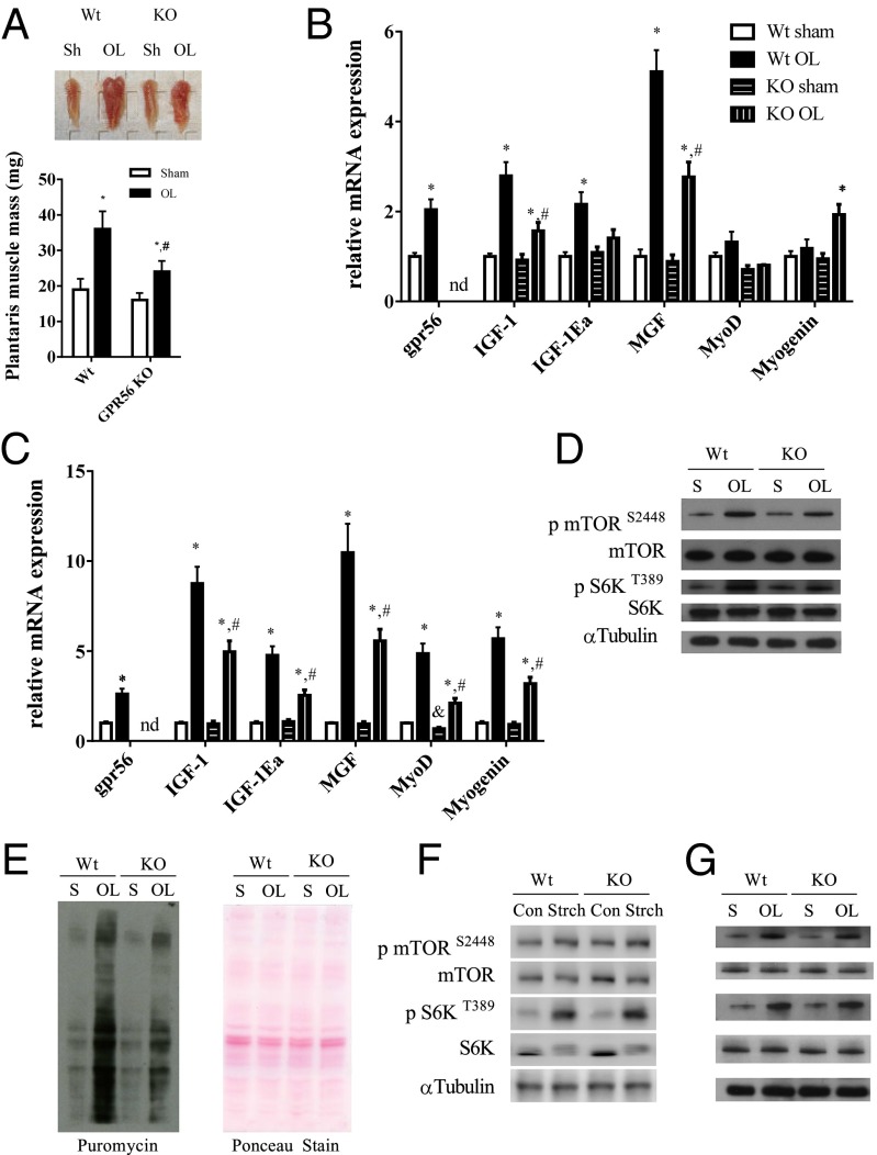
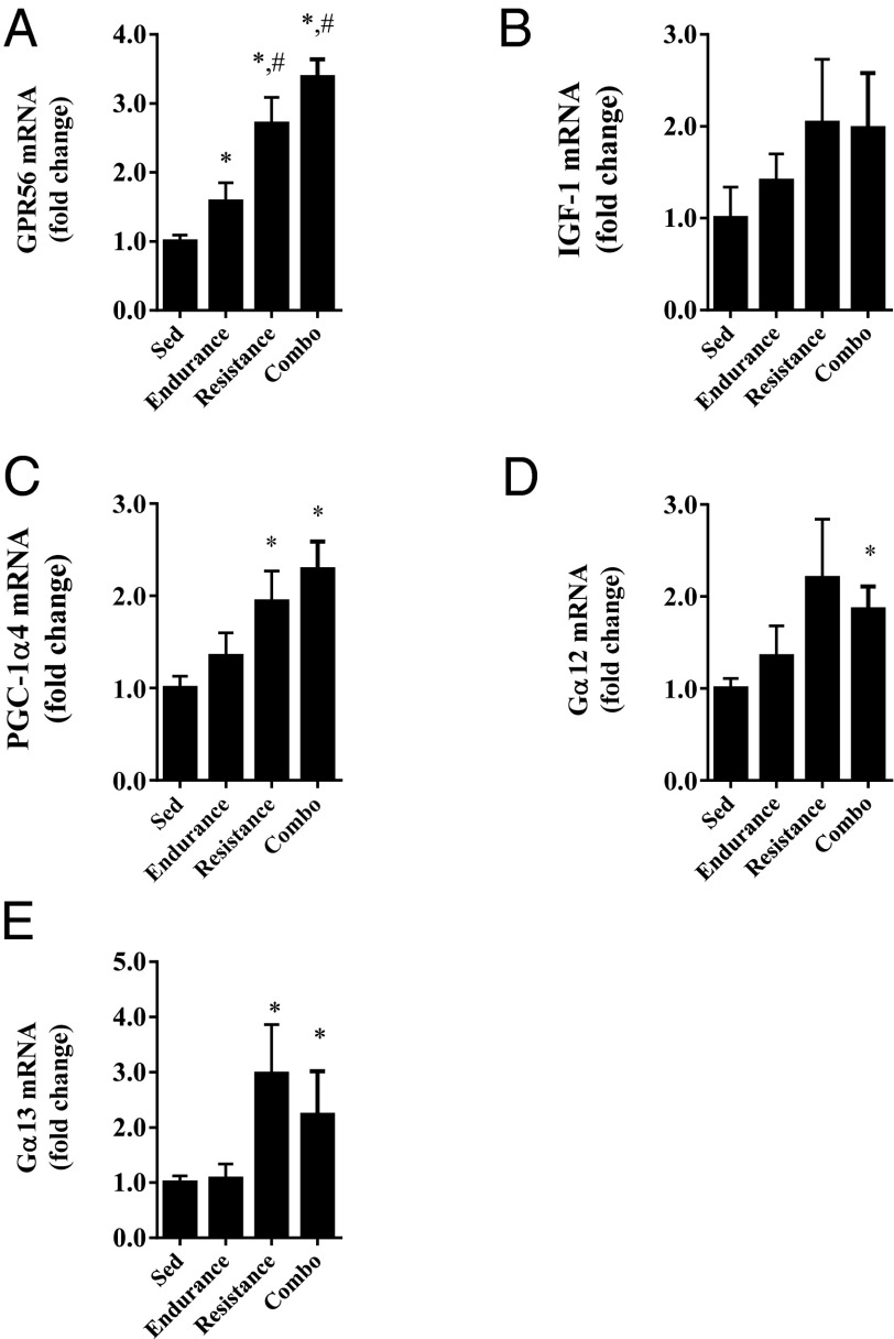
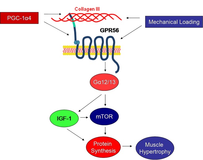
Peroxisome proliferator-activated receptor gamma coactivator 1-alpha 4 (PGC-1α4) is a protein isoform derived by alternative splicing of the PGC1α mRNA and has been shown to promote muscle hypertrophy. We show here that G protein-coupled receptor 56 (GPR56) is a transcriptional target of PGC-1α4 and is induced in humans by resistance exercise. Furthermore, the anabolic effects of PGC-1α4 in cultured murine muscle cells are dependent on GPR56 signaling, because knockdown of GPR56 attenuates PGC-1α4-induced muscle hypertrophy in vitro. Forced expression of GPR56 results in myotube hypertrophy through the expression of insulin-like growth factor 1, which is dependent on Gα12/13 signaling. A murine model of overload-induced muscle hypertrophy is associated with increased expression of both GPR56 and its ligand collagen type III, whereas genetic ablation of GPR56 expression attenuates overload-induced muscle hypertrophy and associated anabolic signaling. These data illustrate a signaling pathway through GPR56 which regulates muscle hypertrophy associated with resistance/loading-type exercise.

Keywords: GPR56; Gα12/13; mTOR; muscle hypertrophy; overload.


Conflict of interest statement

Conflict of interest statement: D.J.G. and Z.W. are employees of, and stockholders in, Novartis.

Figures

Fig. 1.
Fig. 1.
PGC-1α4 drives expression of GPR56 and Col3a1 in primary myotubes. (A and B) GPR56 mRNA (A) and protein (B) expression with adenoviral expression of GFP, PGC-1α1, or PGC-1α4 in primary myotubes. (C and D) GPR56 mRNA (C) and protein (D) expression in muscle from wild-type, Myo PGC-1α4 (−/+), and Myo PGC-1α4 (+/+) mice. (E) Col3a1 mRNA expression with adenoviral expression of GFP, PGC-1α1, or PGC-1α4 in primary myotubes. (F) Peptide expression of collagen III peptides in the media of primary myotubes expressing GFP or PGC-1α4. (G) GPR56 and Col3a1 mRNA expression after intramuscular injection of PGC-1α4 adenovirus or LacZ control. *Denotes significant difference from GFP or wild-type mice. #Denotes significant difference from Myo PGC-1α4 (−/+) mice. Data are presented as mean + SE (n = 3 or 4 per group).
Fig. 2.
Fig. 2.
GPR56 knockdown attenuates PGC-1α4–induced hypertrophy. (A) Myotube morphology with adenoviral (Ad) expression of PGC-1α4 with or without an shRNA against GPR56. (Scale bar, 50 μm.) scr sh, scramble short hairpin. (BD) Myotube diameter (B), myostatin mRNA (C), and IGF1 mRNA (D) expression in myotubes with adenoviral forced expression of PGC-1α4 with or without a short hairpin to GPR56. (E) Representative Western blot of total and phosphorylated mTOR and S6K protein expression. *Denotes significant difference from GFP control myotubes. #Denotes significant difference from myotubes with forced expression of PGC-1α4. Data are presented as mean + SE (n = 3 per group).
Fig. 3.
Fig. 3.
Primary mouse myotubes expressing GPR56 show cellular hypertrophy. (A) Primary mouse myotubes infected with recombinant adenovirus expressing GFP alone with or without collagen III or expressing GPR56 with or without collagen III. (Scale bar, 50 μm.) (B and C) Myotube diameter (B) and IGF1 isoform and myostatin mRNA expression (C) in myotubes expressing GFP or GPR56 with or without collagen III. (D) Representative Western blots of phosphorylated and total mTOR and S6K protein expression in myotubes expressing GFP or GPR56 with or without collagen III. (E and F) Myotube diameter (E) and IGF1 mRNA (F) of myotubes expressing GFP and GPR56 with or without expression of a DN Gα12/13 subunit. (G) Representative Western blots of phosphorylated and total mTOR and S6K protein expression in myotubes expressing GFP and GPR56 with or without expression of a DN Gα12/13 subunit. *Denotes significant difference from GFP control myotubes. Data are presented as mean + SE (n = 3 per group).
Fig. 4.
Fig. 4.
Muscle GPR56 expression is increased in models of muscle hypertrophy. (A) GPR56, PGC-1α1, and PGC-1α4 mRNA expression in mouse muscle after 30 d of free wheel running or sedentary control. (B) Muscle protein expression of GPR56 after 30 d of wheel running (Ex) or sedentary control. (C and D) GPR56 mRNA (C) and protein (D) expression throughout a time course of functional overload (OL). *Denotes significant difference from sedentary or sham controls. (E, Upper) Representative immunohistochemistry of muscle cross-sections stained with wheat germ agglutinin (red) and GFP (green) after 21 d of functional overload. DAPI staining is shown in blue. (Lower) Cross-sectional area of GFP+ and GFP myofibers. (F, Upper) Representative Immunohistochemistry of muscle cross-sectional area stained with wheat germ agglutinin and GFP after 10 d of functional overload. (Lower) Cross-sectional area of GFP+ and GFP myofibers. &Denotes significant difference from GFP myofibers. Data are presented as mean + SE (n = 5–7 per group). The dashed lines across the graphs represent the mean cross-sectional area (CSA) of unloaded sham control muscle for the given experiment. (Scale bars, 50 μm.)
Fig. 5.
Fig. 5.
Loss of GPR56 attenuates overload-induced hypertrophy and anabolic signaling. (A, Upper) Representative plantaris muscles from wild-type and Gpr56-KO mice subjected to either sham treatment or overload. (Lower) Quantified muscle wet weights. (B and C) Plantaris mRNA expression at 21 (B) and 2 (C) d of functional overload. nd, not determined. (D) Representative Western blots of phosphorylated and total mTOR and S6K for wild-type and Gpr56-KO mice subjected to 7 d of overload. (E, Left) Representative Western blot of puromycin-incorporated proteins and (Right) Ponceau staining. (F) Representative Western blots of phosphorylated and total mTOR and S6K of wild-type and Gpr56-KO muscle after 90 min of acute stretching. Con, control; Strch, stretching. (G) Representative Western blots of phosphorylated and total mTOR and S6K of wild-type and Gpr56-KO muscle after 90 min of overload. *Denotes significant difference from sham control. #Denotes significant difference from wild-type overloaded muscle. Data are presented as mean + SE (n = 6 or 7 per group).
Fig. 6.
Fig. 6.
GPR56 and Gα12/13 subunit mRNA are increased in human muscle with resistance-based exercise. GPR56 (A), IGF1 (B), PGC-1a4 (C), Ga12 (D), and Ga13 (E) mRNA expression in human muscle after a 12-wk exercise training in groups undergoing endurance training, resistance training, or a combination of both endurance and resistance training, and in sedentary controls. *Denotes significant difference from sedentary control. #Denotes significant difference from endurance-trained subjects. Data are presented as mean + SE (n = 5–10 per group).
Fig. 7.
Fig. 7.
Working model of GPR56 signaling in skeletal muscle. Driven by PGC-1α4 or independently by mechanical loading, GPR56 signals through the Gα12/13 subunit to induce IGF1 expression and downstream mTOR activation. The activation in mTOR signaling is associated with increased muscle protein synthesis and subsequent hypertrophy.

References

    1. Hornberger TA. Mechanotransduction and the regulation of mTORC1 signaling in skeletal muscle. Int J Biochem Cell Biol. 2011;43(9):1267–1276. - PMC - PubMed
    1. Tesch PA. Skeletal muscle adaptations consequent to long-term heavy resistance exercise. Med Sci Sports Exerc. 1988;20(5) Suppl:S132–S134. - PubMed
    1. Goldberg AL. Protein synthesis during work-induced growth of skeletal muscle. J Cell Biol. 1968;36(3):653–658. - PMC - PubMed
    1. Hunter GR, McCarthy JP, Bamman MM. Effects of resistance training on older adults. Sports Med. 2004;34(5):329–348. - PubMed
    1. Ruas JL, et al. A PGC-1α isoform induced by resistance training regulates skeletal muscle hypertrophy. Cell. 2012;151(6):1319–1331. - PMC - PubMed

Publication types

MeSH terms

Substances