Middle fossa arachnoid cysts and inner ear symptoms: Are they related?
- PMID: 25336883
- PMCID: PMC4201406
Middle fossa arachnoid cysts and inner ear symptoms: Are they related?
Abstract
Background: Arachnoid cysts most frequently occur in the middle cranial fossa and when they are symptomatic, patients present with central nervous symptoms. Nevertheless, a large proportion of arachnoid cysts are incidentally diagnosed during neuroimaging in cases with nonspecific symptoms.
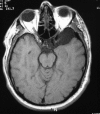
Report of cases: The cases of two males with middle cranial fossa arachnoid cysts with nonspecific inner ear symptoms were retrospectively reviewed. The first patient presented with mild headache, nausea, vertigo, unsteadiness, and tinnitus on the left ear while the second patient's main complaint was left sided tinnitus. Both patients (initially managed for peripheral disorders) underwent a thorough clinical and electrophysiological evaluation. Because of the patients' persistent clinical symptoms, and indications of CNS disorder in the first case, neuroimaging by brain MRI was performed revealing a middle cranial fossa arachnoid cyst in both patients.
Conclusion: Occasionally, patients with arachnoid cysts may present with mild, atypical or intermittent and irrelevant symptoms which can mislead diagnosis. Otorhinolaryngologists should be aware of the fact that atypical, recurrent or intermittent symptoms may masquerade a CNS disorder. Hippokratia 2014; 18 (2):168-171.
Keywords: arachnoid cyst; atypical presenting symptoms; hearing loss; middle cranial fossa; tinnitus; vertigo.
Figures


References
-
- Pradilla G, Jallo G. Arachnoid cysts: case series and review of the literature. Neurosurg Focus. 2007;22:E7. - PubMed
-
- Galassi E, Tognetti F, Gaist G, Fagioli L, Frank F, Frank G. CT scan and metrizamide CT cisternography in arachnoid cyst of middle cranial fossa: classification and pathophysiological aspect. Surg Neurol. 1982;17:363–369. - PubMed
-
- Greenberg MS. Handbook of Neurosurgery. 6th ed. Thieme Medical Publishers, New York. 2006
-
- Harsh G 4th, Edwards MS, Wilson CB. Intracranial arachnoid cysts in children. J Neurosurg. 1986;64:835–842. - PubMed
Publication types
LinkOut - more resources
Full Text Sources
Research Materials
