

Morphological differences in skeletal muscle atrophy of rats with motor nerve and/or sensory nerve injury
- PMID: 25337102
- PMCID: PMC4200706
- DOI: 10.3969/j.issn.1673-5374.2012.32.004
Morphological differences in skeletal muscle atrophy of rats with motor nerve and/or sensory nerve injury
Abstract
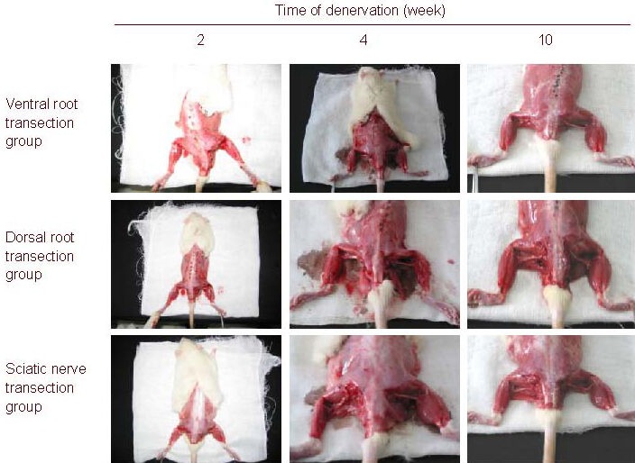
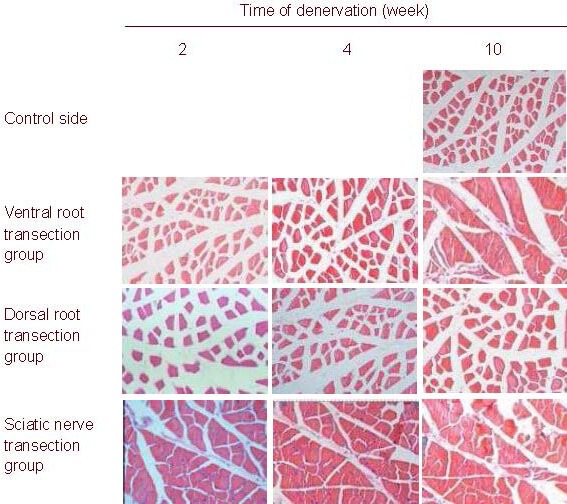
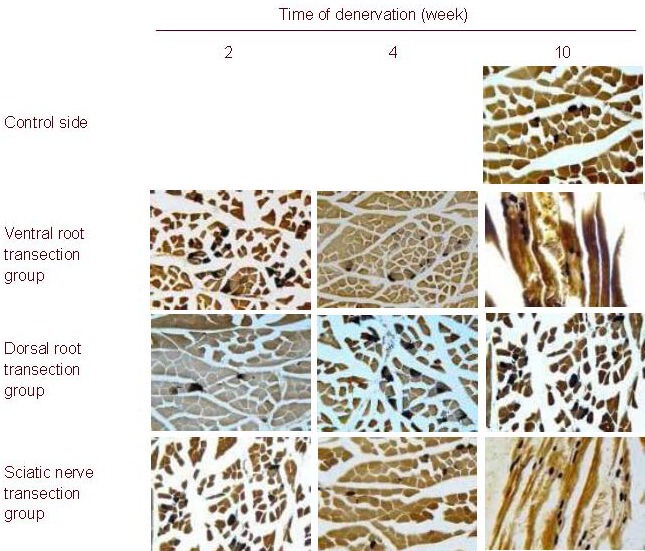
Skeletal muscle atrophy occurs after denervation. The present study dissected the rat left ventral root and dorsal root at L4-6 or the sciatic nerve to establish a model of simple motor nerve injury, sensory nerve injury or mixed nerve injury. Results showed that with prolonged denervation time, rats with simple motor nerve injury, sensory nerve injury or mixed nerve injury exhibited abnormal behavior, reduced wet weight of the left gastrocnemius muscle, decreased diameter and cross-sectional area and altered ultrastructure of muscle cells, as well as decreased cross-sectional area and increased gray scale of the gastrocnemius muscle motor end plate. Moreover, at the same time point, the pathological changes were most severe in mixed nerve injury, followed by simple motor nerve injury, and the changes in simple sensory nerve injury were the mildest. These findings indicate that normal skeletal muscle morphology is maintained by intact innervation. Motor nerve injury resulted in larger damage to skeletal muscle and more severe atrophy than sensory nerve injury. Thus, reconstruction of motor nerves should be considered first in the clinical treatment of skeletal muscle atrophy caused by denervation.
Keywords: motor end plate; muscular atrophy; neural regeneration; rats; simple nerve injury; ultrastructure.
Conflict of interest statement
Figures

Similar articles
-
Motor denervation induces altered muscle fibre type densities and atrophy in a rat model of neuropathic pain.Neurosci Lett. 1998 May 15;247(2-3):204-8. doi: 10.1016/s0304-3940(98)00304-8. Neurosci Lett. 1998. PMID: 9655629
-
Third-degree hindpaw burn injury induced apoptosis of lumbar spinal cord ventral horn motor neurons and sciatic nerve and muscle atrophy in rats.Biomed Res Int. 2015;2015:372819. doi: 10.1155/2015/372819. Epub 2015 Jan 28. Biomed Res Int. 2015. PMID: 25695065 Free PMC article.
-
Calpain 3 Expression Pattern during Gastrocnemius Muscle Atrophy and Regeneration Following Sciatic Nerve Injury in Rats.Int J Mol Sci. 2015 Nov 11;16(11):26927-35. doi: 10.3390/ijms161126003. Int J Mol Sci. 2015. PMID: 26569227 Free PMC article.
-
Skeletal muscle injury and repair: the effect of disuse and denervation on muscle and clinical relevance in pedicled and free muscle flaps.J Reconstr Microsurg. 2012 Nov;28(9):581-7. doi: 10.1055/s-0032-1315784. Epub 2012 Jun 18. J Reconstr Microsurg. 2012. PMID: 22711205 Review.
-
Chapter 25: Phototherapy in peripheral nerve injury: effects on muscle preservation and nerve regeneration.Int Rev Neurobiol. 2009;87:445-64. doi: 10.1016/S0074-7742(09)87025-6. Int Rev Neurobiol. 2009. PMID: 19682654 Review.
Cited by
-
Neuromuscular Dysfunction Precedes Cognitive Impairment in a Mouse Model of Alzheimer's Disease.Function (Oxf). 2023 Dec 4;5(1):zqad066. doi: 10.1093/function/zqad066. eCollection 2024. Function (Oxf). 2023. PMID: 38111538 Free PMC article.
-
Edaravone promotes functional recovery after mechanical peripheral nerve injury.Neural Regen Res. 2014 Sep 15;9(18):1709-15. doi: 10.4103/1673-5374.141808. Neural Regen Res. 2014. PMID: 25374594 Free PMC article.
-
The neurochemistry of peripheral nerve regeneration.Indian J Plast Surg. 2017 Jan-Apr;50(1):5-15. doi: 10.4103/ijps.IJPS_14_17. Indian J Plast Surg. 2017. PMID: 28615804 Free PMC article.
-
Effect of melatonin supplemented at the light or dark period on recovery of sciatic nerve injury in rats.EXCLI J. 2017 Mar 6;16:138-150. doi: 10.17179/excli2016-763. eCollection 2017. EXCLI J. 2017. PMID: 28435433 Free PMC article.
-
Voluntary Exercise Training: Analysis of Mice in Uninjured, Inflammatory, and Nerve-Injured Pain States.PLoS One. 2015 Jul 21;10(7):e0133191. doi: 10.1371/journal.pone.0133191. eCollection 2015. PLoS One. 2015. PMID: 26196858 Free PMC article.
References
-
- Tews DS, Goebel HH, Schneider I, et al. DNA- fragmentation and expression of apoptosis-related proteins in experimentally denervated and reinnervated rat facial muscle. Neuropathol Appl Neurobiol. 1997;23(2):141–149. - PubMed
-
- Li ZB, Lehar M, Samlan R, et al. Proteomic analysis of rat laryngeal muscle following denervation. Proteomics. 2005;5(18):4764–4776. - PubMed
-
- Hart AM, Terenghi G, Wiberg M. Neuronal death after peripheral nerve injury and experimental strategies for neuroprotection. Neurol Res. 2008;30(10):999–1011. - PubMed
LinkOut - more resources
Full Text Sources