

Promotion of initial anti-tumor effect via polydopamine modified doxorubicin-loaded electrospun fibrous membranes
- PMID: 25337186
- PMCID: PMC4203157
Promotion of initial anti-tumor effect via polydopamine modified doxorubicin-loaded electrospun fibrous membranes
Abstract
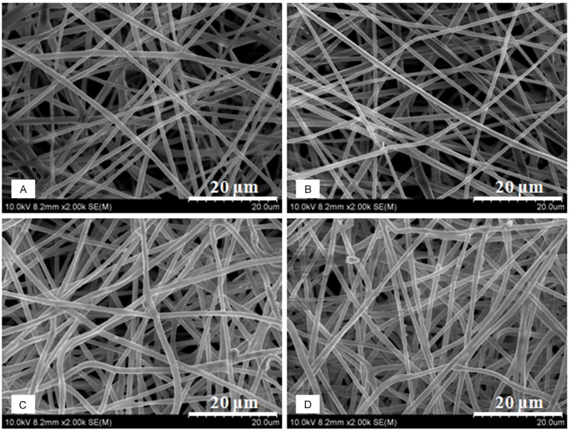
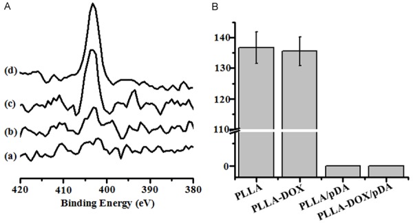
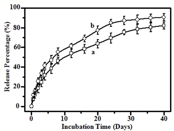
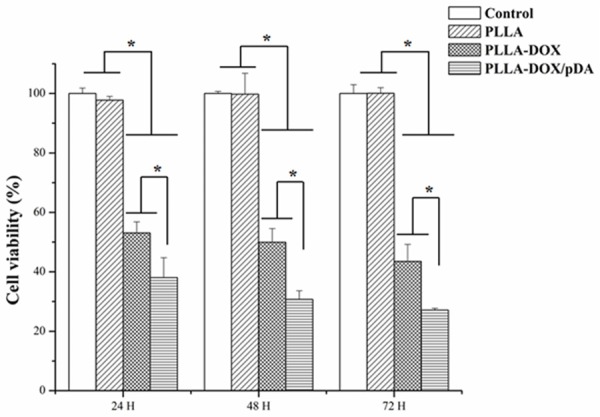
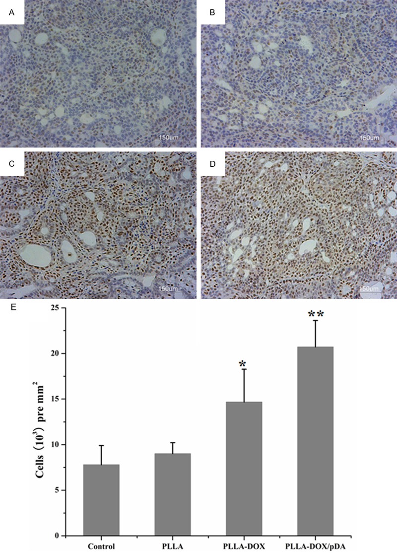
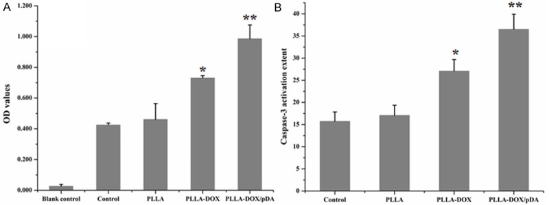
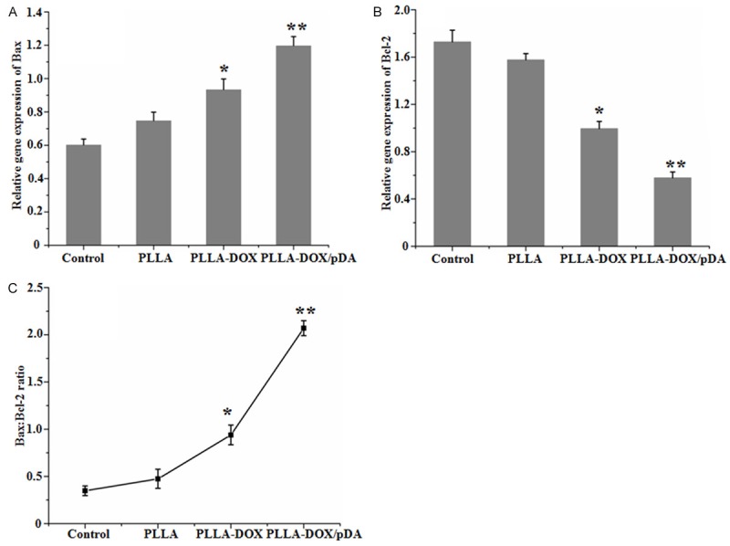
Drug-loaded electrospun PLLA membranes are not conducive to adhesion between materials and tissues due to the strong hydrophobicity of PLLA, which possibly attenuate the drugs' effect loaded on the materials. In the present work, we developed a facile method to improve the hydrophilicity of doxorubicin (DOX)-loaded electrospun PLLA fibrous membranes, which could enhance the anti-tumor effect at the early stage after implantation. A mussel protein, polydopamine (PDA), could be easily grafted on the surface of hydrophobic DOX-loaded electrospun PLLA membranes (PLLA-DOX/pDA) in water solution. The morphology analysis of PLLA-DOX/pDA fibers displayed that though the fiber diameter was slightly swollen, they still maintained a 3D fibrous structure, and the XPS analysis certified that pDA had successfully been grafted onto the surface of the fibers. The results of surface wettability analysis showed that the contact angle decreased from 136.7° to 0° after grafting. In vitro MTT assay showed that the cytotoxicity of PLLA-DOX/pDA fibers was the strongest, and the stereologic cell counting assay demonstrated that the adhesiveness of PLLA/pDA fiber was significantly better than PLLA fiber. In vivo tumor-bearing mice displayed that, after one week of implantation, the tumor apoptosis and necrosis of PLLA-DOX/pDA fibers were the most obvious from histopathology and TUNEL assay. The caspase-3 activity of PLLA-DOX/pDA group was the highest using biochemical techniques, and the Bax: Bcl-2 ratio increased significantly in PLLA-DOX/pDA group through qRT-PCR analysis. All the results demonstrated that pDA can improve the affinity of the electrospun PLLA membranes and enhance the drug effect on tumors.
Keywords: Cancer; PLLA; electrospun; hydrophobicity; polydopamine.
Figures

Similar articles
-
Mn2+-coordinated PDA@DOX/PLGA nanoparticles as a smart theranostic agent for synergistic chemo-photothermal tumor therapy.Int J Nanomedicine. 2017 Apr 24;12:3331-3345. doi: 10.2147/IJN.S132270. eCollection 2017. Int J Nanomedicine. 2017. PMID: 28479854 Free PMC article.
-
Self-coated interfacial layer at organic/inorganic phase for temporally controlling dual-drug delivery from electrospun fibers.Colloids Surf B Biointerfaces. 2015 Jun 1;130:1-9. doi: 10.1016/j.colsurfb.2015.03.058. Epub 2015 Apr 3. Colloids Surf B Biointerfaces. 2015. PMID: 25879640
-
Polydopamine-Based Nanoparticles for Synergistic Chemotherapy of Prostate Cancer.Int J Nanomedicine. 2024 Jul 3;19:6717-6730. doi: 10.2147/IJN.S468946. eCollection 2024. Int J Nanomedicine. 2024. PMID: 38979530 Free PMC article.
-
Amplified Photoacoustic Signal and Enhanced Photothermal Conversion of Polydopamine-Coated Gold Nanobipyramids for Phototheranostics and Synergistic Chemotherapy.ACS Appl Mater Interfaces. 2020 Apr 1;12(13):14866-14875. doi: 10.1021/acsami.9b22979. Epub 2020 Mar 19. ACS Appl Mater Interfaces. 2020. PMID: 32153178
-
Combined Chemo- and Photothermal Therapies of Non-Small Cell Lung Cancer Using Polydopamine/Au Hollow Nanospheres Loaded with Doxorubicin.Int J Nanomedicine. 2024 Sep 14;19:9597-9612. doi: 10.2147/IJN.S473137. eCollection 2024. Int J Nanomedicine. 2024. PMID: 39296938 Free PMC article.
Cited by
-
Multifunctional Electrospun Nanofibers for Enhancing Localized Cancer Treatment.Small. 2018 Jun 27:e1801183. doi: 10.1002/smll.201801183. Online ahead of print. Small. 2018. PMID: 29952070 Free PMC article. Review.
References
-
- Sakthivel KM, Guruvayoorappan C. Acacia ferruginea inhibits tumor progression by regulating inflammatory mediators-(TNF-a, iNOS, COX-2, IL-1β, IL-6, IFN-γ, IL-2, GM-CSF) and pro-angiogenic growth factor-VEGF. Asian Pac J Cancer Preve. 2013;14:3909–3919. - PubMed
Publication types
MeSH terms
Substances
LinkOut - more resources
Full Text Sources
Medical
Research Materials