A fine decision tree consisted of CK5/6, IMP3 and TTF1 for cytological diagnosis among reactive mesothelial cells, metastatic adenocarcinoma of lung and non-lung origin in pleural effusion
- PMID: 25337222
- PMCID: PMC4203193
A fine decision tree consisted of CK5/6, IMP3 and TTF1 for cytological diagnosis among reactive mesothelial cells, metastatic adenocarcinoma of lung and non-lung origin in pleural effusion
Abstract
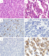
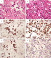
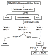
The utility of combination with CK5/6, IMP3 and TTF1 to differentiate among reactive mesothelial cells (RMs), metastatic adenocarcinoma of lung (LAC) and non-lung (NLAC) origin was investigated by using immunocytochemistry (ICC) and conventional PCR (C-PCR) in pleural effusion. A total of 108 cell blocks (32 RMs, 51 LAC and 25 NLAC were evaluated by ICC for CK5/6, IMP3 and TTF1 protein expression. In addition, we further performed C-PCR for amplification of CK5/6, IMP3 and TTF1 DNA from 28 specimens (9 MAC and 7 RMs, 6 LAC and 6 NLAC) for molecular diagnosis. CK5/6 staining was observed in the majority of reactive specimens (78.1%) and was rare in adenocarcinoma cells (14.5%), whereas the opposite was true for IMP3 and TTF1. We found a high frequency of TTF1 positivity (76.5%) in LAC, but not in NLAC (4.0%); while there was no significant difference of IMP3 expression in LAC (88.2%) and NLAC (88.0%). The 487 bp DNA fragments of IMP3 was expected to be amplified in 6/9 of adenocarcinoma cases showed negative in ICC; and the 394 bp DNA fragments of CK5/6 was also expected to be amplified in 4/7 of RMs cases showed negative in ICC. Consistent with ICC results, there was significant difference of TTF1 expression in the LAC and NLAC compared with IMP3 expression. The combination with CK5/6, IMP3 and TTF1 immunostaining appears to be useful to improve the accuracy of cytological diagnoses between RMs, metastatic adenocarcinoma of lung and non-lung origin in pleural effusion. In addition, C-PCR may act as a useful supplemental approach for ICC, especially negative cases in ICC for differential cytological diagnosis.
Keywords: CK5/6; IMP3; PCR; TTF1; adenocarcinoma; pleural effusion.
Figures


Similar articles
-
Utility of anti-L523S antibody in the diagnosis of benign and malignant serous effusions.Cancer. 2008 Feb 25;114(1):49-56. doi: 10.1002/cncr.23254. Cancer. 2008. PMID: 18098206
-
IMP3/L523S, a novel immunocytochemical marker that distinguishes benign and malignant cells: the expression profiles of IMP3/L523S in effusion cytology.Hum Pathol. 2010 May;41(5):745-50. doi: 10.1016/j.humpath.2009.04.030. Epub 2010 Jan 13. Hum Pathol. 2010. PMID: 20060157
-
Diagnostic usefulness of EMA, IMP3, and GLUT-1 for the immunocytochemical distinction of malignant cells from reactive mesothelial cells in effusion cytology using cytospin preparations.Diagn Cytopathol. 2011 Jun;39(6):395-401. doi: 10.1002/dc.21398. Epub 2010 May 27. Diagn Cytopathol. 2011. PMID: 21574259
-
Ber-EP4 staining in effusion cytology: A potential source of false positives.Rev Esp Patol. 2021 Apr-Jun;54(2):114-122. doi: 10.1016/j.patol.2020.04.005. Epub 2020 Jul 18. Rev Esp Patol. 2021. PMID: 33726887 Review.
-
Adenocarcinoma cells in effusion cytology as a diagnostic pitfall with potential impact on clinical management: a case report with brief review of immunomarkers.Diagn Cytopathol. 2014 Mar;42(3):253-8. doi: 10.1002/dc.22915. Epub 2012 Nov 16. Diagn Cytopathol. 2014. PMID: 23161830 Free PMC article. Review.
Cited by
-
Surgical intervention for non-small-cell lung cancer with minimal malignant pleural effusion.Surg Today. 2023 Jun;53(6):655-662. doi: 10.1007/s00595-022-02606-4. Epub 2022 Oct 31. Surg Today. 2023. PMID: 36310332
-
Is cell block technique useful to predict histological type in patients with ovarian mass and/or body cavity fluids?Nagoya J Med Sci. 2020 May;82(2):225-235. doi: 10.18999/nagjms.82.2.225. Nagoya J Med Sci. 2020. PMID: 32581403 Free PMC article.
-
Papillary Thyroid Carcinoma Cervical Lymph Node Metastasis with Cystic Change Differentiated from Congenital Cystic Lesions with the Assistance of Immunohistochemistry: A Case Study.Head Neck Pathol. 2017 Sep;11(3):301-305. doi: 10.1007/s12105-016-0762-1. Epub 2016 Oct 21. Head Neck Pathol. 2017. PMID: 27770399 Free PMC article.
-
Diagnostic value of thyroid transcription factor-1 for pleural or other serous metastases of pulmonary adenocarcinoma: a meta-analysis.Sci Rep. 2016 Jan 25;6:19785. doi: 10.1038/srep19785. Sci Rep. 2016. PMID: 26806377 Free PMC article.
References
-
- Heffner JE. Diagnosis and management of malignant pleural effusions. Respirology. 2008;13:5–20. - PubMed
-
- Hsieh TC, Huang WW, Lai CL, Tsao SM, Su CC. Diagnostic value of tumor markers in lung adenocarcinoma-associated cytologically negative pleural effusions. Cancer Cytopathol. 2013;121:483–8. - PubMed
-
- Metzgeroth G, Kuhn C, Schultheis B, Hehlmann R, Hastka J. Diagnostic accuracy of cytology and immunocytology in carcinomatous effusions. Cytopathology. 2007;19:205–211. - PubMed
-
- Kim JH, Kim GE, Choi YD, Lee JS, Lee JH, Nam JH, Choi C. Immunocytochemical panel for distinguishing between adenocarcinomas and reactive mesothelial cells in effusion cell blocks. Diagn Cytopathol. 2009;37:258–61. - PubMed
Publication types
MeSH terms
Substances
LinkOut - more resources
Full Text Sources
Medical
Research Materials
Miscellaneous
