

Parathyroid hormone induces epithelial-to-mesenchymal transition via the Wnt/β-catenin signaling pathway in human renal proximal tubular cells
- PMID: 25337242
- PMCID: PMC4203213
Parathyroid hormone induces epithelial-to-mesenchymal transition via the Wnt/β-catenin signaling pathway in human renal proximal tubular cells
Abstract
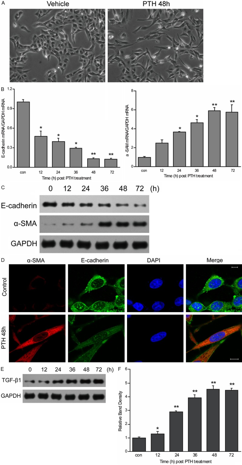
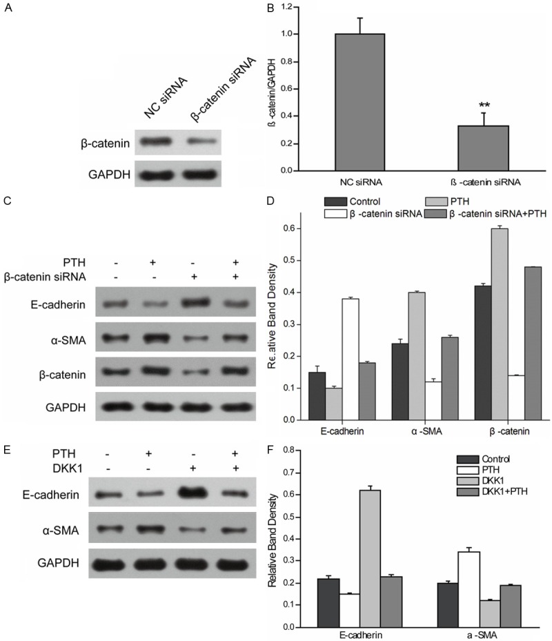
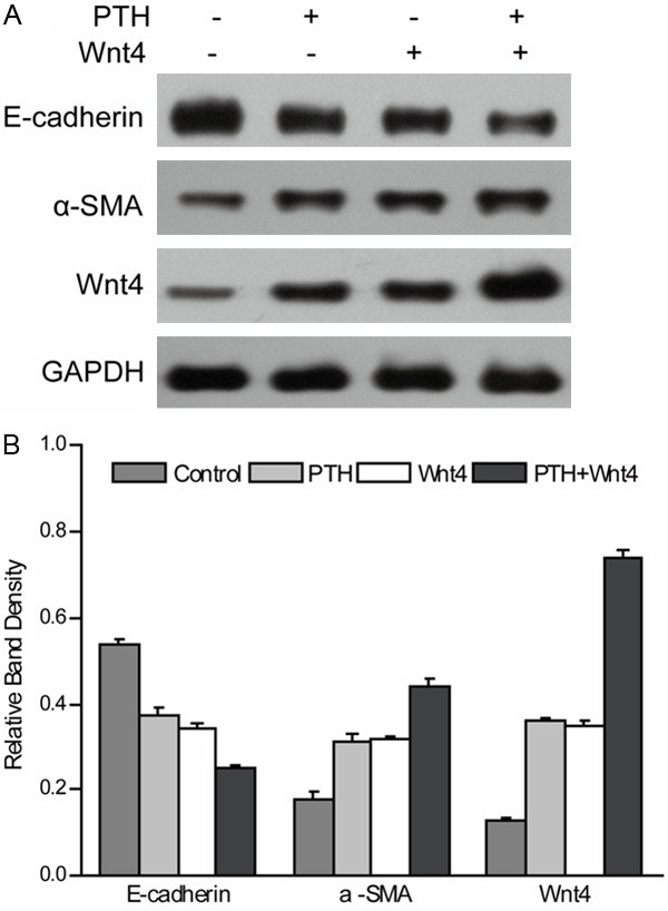
Epithelial-to-mesenchymal transition (EMT) has been shown to play an important role in renal fibrogenesis. Recent studies suggested parathyroid hormone (PTH) could accelerate EMT and subsequent organ fibrosis. However, the precise molecular mechanisms underlying PTH-induced EMT remain unknown. The present study was to investigate whether Wnt/β-catenin signaling pathway is involved in PTH-induced EMT in human renal proximal tubular cells (HK-2 cells) and to determine the profile of gene expression associated with PTH-induced EMT. PTH could induce morphological changes and gene expression characteristic of EMT in cultured HK-2 cells. Suppressing β-catenin expression or DKK1 limited gene expression characteristic of PTH-induced EMT. Based on the PCR array analysis, PTH treatment resulted in the up-regulation of 18 genes and down-regulation of 9 genes compared with the control. The results were further supported by a western blot analysis, which showed the increased Wnt4 protein expression. Wnt4 overexpression also promotes PTH-induced EMT in HK-2 cells. The findings demonstrated that PTH-induced EMT in HK-2 cells is mediated by Wnt/β-catenin signal pathway, and Wnt4 might be a key gene during PTH-induced EMT.
Keywords: Epithelial-to-mesenchymal transition; Wnt/β-catenin signal pathway; parathyroid hormone; renal tubular epithelial cell.
Figures
Similar articles
-
Connective tissue growth factor induces tubular epithelial to mesenchymal transition through the activation of canonical Wnt signaling in vitro.Ren Fail. 2015 Feb;37(1):129-35. doi: 10.3109/0886022X.2014.967699. Epub 2014 Oct 8. Ren Fail. 2015. PMID: 25296105
-
FKN Facilitates HK-2 Cell EMT and Tubulointerstitial Lesions via the Wnt/β-Catenin Pathway in a Murine Model of Lupus Nephritis.Front Immunol. 2019 Apr 30;10:784. doi: 10.3389/fimmu.2019.00784. eCollection 2019. Front Immunol. 2019. PMID: 31134047 Free PMC article.
-
lncRNA MALAT1 mediated high glucose-induced HK-2 cell epithelial-to-mesenchymal transition and injury.J Physiol Biochem. 2019 Nov;75(4):443-452. doi: 10.1007/s13105-019-00688-2. Epub 2019 Aug 6. J Physiol Biochem. 2019. PMID: 31388927
-
Emerging Therapeutic Strategies for Attenuating Tubular EMT and Kidney Fibrosis by Targeting Wnt/β-Catenin Signaling.Front Pharmacol. 2022 Jan 10;12:830340. doi: 10.3389/fphar.2021.830340. eCollection 2021. Front Pharmacol. 2022. PMID: 35082683 Free PMC article. Review.
-
Modulation of Epithelial to Mesenchymal Transition Signaling Pathways by Olea Europaea and Its Active Compounds.Int J Mol Sci. 2019 Jul 16;20(14):3492. doi: 10.3390/ijms20143492. Int J Mol Sci. 2019. PMID: 31315241 Free PMC article. Review.
Cited by
-
Acidosis-induced p38-kinase activation triggers an IL-6-mediated crosstalk of renal proximal tubule cells with fibroblasts leading to their inflammatory response.Cell Commun Signal. 2025 Apr 11;23(1):180. doi: 10.1186/s12964-025-02180-5. Cell Commun Signal. 2025. PMID: 40217316 Free PMC article.
-
Origin of cells and network information.World J Stem Cells. 2015 Apr 26;7(3):535-40. doi: 10.4252/wjsc.v7.i3.535. World J Stem Cells. 2015. PMID: 25914760 Free PMC article.
-
Induced Neurodifferentiation of hBM-MSCs through Activation of the ERK/CREB Pathway via Pulsed Electromagnetic Fields and Physical Stimulation Promotes Neurogenesis in Cerebral Ischemic Models.Int J Mol Sci. 2022 Jan 21;23(3):1177. doi: 10.3390/ijms23031177. Int J Mol Sci. 2022. PMID: 35163096 Free PMC article.
-
[Expression of kinesin KIF3A in the kidney of mice with unilateral ureteral obstruction].Nan Fang Yi Ke Da Xue Xue Bao. 2020 Feb 29;40(2):219-224. doi: 10.12122/j.issn.1673-4254.2020.02.13. Nan Fang Yi Ke Da Xue Xue Bao. 2020. PMID: 32376524 Free PMC article. Chinese.
-
Baicalin ameliorates renal fibrosis via inhibition of transforming growth factor β1 production and downstream signal transduction.Mol Med Rep. 2017 Apr;15(4):1702-1712. doi: 10.3892/mmr.2017.6208. Epub 2017 Feb 16. Mol Med Rep. 2017. PMID: 28260014 Free PMC article.
References
-
- Noronha IL, Fujihara CK, Zatz R. The inflammatory component in progressive renal disease - are interventions possible? Nephrol Dial Transplant. 2002;17:363–368. - PubMed
-
- Fujihara CK, Malheiros DM, Zatz R, Noronha IL. Mycophenolate mofetil attenuates renal injury in the rat remnant kidney. Kidney Int. 1998;54:1510–1519. - PubMed
-
- Lewis EJ, Hunsicker LG, Clarke WR, Berl T, Pohl MA, Lewis JB, Ritz E, Atkins RC, Rohde R, Raz I Collaborative Study Group. Collaborative Study Group. Renoprotective effect of the angiotensin-receptor antagonist irbesartan in patients with nephropathy due to type 2 diabetes. N Engl J Med. 2001;345:851–860. - PubMed
-
- Brenner BM, Cooper ME, de Zeeuw D, Keane WF, Mitch WE, Parving HH, Remuzzi G, Snapinn SM, Zhang Z, Shahinfar S RENAAL Study Investigators. RENAAL Study Investigators. Effects of losartan on renal and cardiovascular outcomes in patients with type 2 diabetes and nephropathy. N Engl J Med. 2001;345:861–869. - PubMed
-
- Carew RW, Wang B, Kantharidis P. The role of EMT in renal fibrosis. Cell Tissue Res. 2012;347:103–116. - PubMed
Publication types
MeSH terms
Substances
LinkOut - more resources
Full Text Sources
Miscellaneous