Review
. 2014 Aug 15;7(9):6323-32.
eCollection 2014.

Primary gliosarcoma with long-survival: report of two cases and review of literature

Affiliations
Review

Primary gliosarcoma with long-survival: report of two cases and review of literature

Zhen Huo et al. Int J Clin Exp Pathol. .

Abstract

Background: Gliosarcoma (GS) is a rare high-grade malignant tumor with poor prognosis. The survival period of GS ranges from 4 to 18.5 months. Rarely would it be over 40 months. Survival of intraventricular GS is less than 8 months.

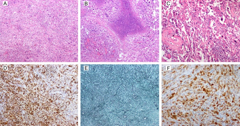
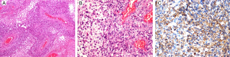
Methods: There were 2 cases of primary gliosarcoma in our hospital with long-term survival after resection, with one of pure intraventricular origin. We confirmed that our diagnosis was correct by light microscopy, GFAP immunohistochemistry and histochemistry of reticular fiber staining.

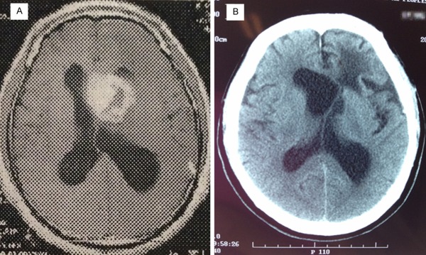
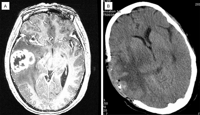
Results: In the first case, a 47-year-old man with intraventricular gliosarcoma survived for 130 months after surgery. In another case, a 63-year-old woman survived for 4 years after resection. Both cases of GS exhibited biphasic glioblastoma and fibrosarcoma with necrosis. According to the review of surgical records, complete tumor resections, including extended resections were carried out in both cases. The two patients received postoperative radiation therapy and chemotherapy without any further recurrence and metastasis.

Conclusions: We reported two cases of GS with long survival. The presented cases demonstrate that, in rare instances, gliosarcoma may show prolonged survival with after surgical excision combined with radiotherapy and chemotherapy.

Keywords: Primary gliosarcoma; intraventricular gliosarcoma; long survival.


Figures

Figure 1
Figure 1
MRI scan of the tumor in Case 1 in 2003 demonstrates an irregular and uneven ring-shaped heterogeneous enhanced lesions around front left lateral ventricle, right shift of brain midline due to pressure (A: axial T-1 contrast-enhanced images). CT scan in 2014 revealed irregular structure in the anterior horn of the left lateralventricle, left shift of brain midline and a large cystic low-density areasin the anterior horn of the left lateral ventricle (B: axial plane).
Figure 2
Figure 2
Section of the tumor in Case 1 shows a mix of fusiform glial and sarcomatous areas and the glial components are split into pieces with distinct sarcomatous cells (A: H&E, original ×40). Another area shows irregular necrosis (B: H&E, original ×40) and significant cellular atypia, mitosis and visible pathological mitosis (C: H&E, original ×150). Sections with GFAP (D: GFAP, ×150) and reticulin (E: Reticulin, ×150) show the GFAP-positive, reticulin-poor glial areas contrasting with GFAP-negative, reticulin-rich sarcomatous components. Both areas are uniformly positive for P16 (F: P16, ×150).
Figure 3
Figure 3
Preoperative MRI scan of the tumor in case 2 reveals a lobulated enhanced lesion in right temporal lobe, cystic area in middle and large edema around lesions (A: axial T-1 contrast-enhanced images). Postoperative CT scan displayed defections in the right temporal lobe partial skull, no residual lesions and a huge area of edema in original brain lesions (B: axial plane).
Figure 4
Figure 4
Section of the tumor in Case 2 showing spindle-shaped sarcomatous components grow around the thick-walled vessel; outside the sarcomatous cells is glial components (A: H&E, original ×40). The glial cells have gemistocytic morphology (B: H&E, original ×300). Immunohistochemical staining for EGFR demonstrates positivity in tumor cells (C: EGFR, ×300).

Similar articles

Cited by

References

    1. Louis DN, Ohgaki H, Wiestler OD, Cavenee WK. WHO classification of tumours of the central nervous system. 4th edition. Lyon: IARC; 2007. Gliosarcoma; pp. 48–49.
    1. Stroebe H. Ueber Entstehung und Bauder Gehirnglioma. Beitr Pathol Anat Allg Pathol. 1895;19:405–486.
    1. Feigin IH, Gross SW. Sarcoma arising in glioblastoma of the brain. Am J Pathol. 1955;31:633–653. - PMC - PubMed
    1. Feigin I, Allen LB, Lipkin L, Gross SW. The endothelial hyperplasia of the cerebral blood vessels with brain tumors, and its sarcomatous transformation. Cancer. 1958;11:264–277. - PubMed
    1. Reis RM, Könü-Lebleblicioglu D, Lopes JM, Kleihues P, Ohgaki H. Genetic profile of gliosarcomas. Am J Pathol. 2000;156:425–432. - PMC - PubMed

MeSH terms

Substances

LinkOut - more resources