

Clinicopathological features of an ascending colon mixed adenoneuroendocrine carcinoma with clinical serosal invasion
- PMID: 25337298
- PMCID: PMC4203269
Clinicopathological features of an ascending colon mixed adenoneuroendocrine carcinoma with clinical serosal invasion
Abstract
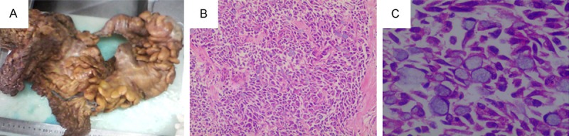
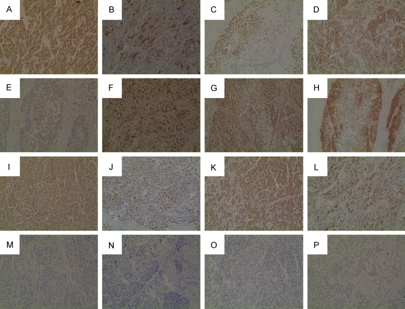
Mixed adenoneuroendocrine carcinoma (MANEC) is exceedingly rare with a poor outcome. In this article, we reported a MANEC in a 68-year-old woman with a symptom of abdominal pain and distension. MANEC derived from the ascending colon with highly aggressive behavior. The diagnosis and distinguish of MANEC must base on histological findings and immunohistochemical findings. In this case, microscopic observation showed tumor cells were arranged in conglobate and nested by fibrous tissue with a visible cell atypia and mitotic. NEC-like and exocrine glandular cells were also been seen in a single neoplasm. MANEC tissues were immunopositive for CK, CK20, P53, CK7, CDX-2, Ki-67 (70%+), E-cad, CD56, CEA, Syn, villin and CgA, and immunonegative for CA125, NSE, ER and PR. Here, the patient was treated by surgical operation and was followed-up near 3 months, no local recurrence and distant metastasis.
Keywords: Mixed adenoneuroendocrine carcinoma; ascending colon; immunohistochemistry.
Figures
References
-
- Scoazec JY, Couvelard A pour le réseau TENpath (réseau national d’expertise pour le diagnostic anatomopathologique des tumeurs neuroendocrines malignes de l’adulte, sporadiques et familiales) [The new WHO classification of digestive neuroendocrine tumors] Ann Pathol. 2011;31:88–92. - PubMed
-
- Clark OH, Benson AR, Berlin JD, Choti MA, Doherty GM, Engstrom PF, Gibbs JF, Heslin MJ, Kessinger A, Kulke MH Kvols L, Salem R, Saltz L, Shah MH, Shibata S, Strosberg JR, Yao JC NCCN Neuroendocrine Tumos Panel Members. NCCN Clinical Practice Guidelines in Oncology: neuroendocrine tumors. J Natl Compr Canc Netw. 2009;7:712–747. - PubMed
Publication types
MeSH terms
Substances
LinkOut - more resources
Full Text Sources
Research Materials
Miscellaneous