

Functional Interaction between the Shell Sub-Region of the Nucleus Accumbens and the Ventral Tegmental Area in Response to Morphine: an Electrophysiological Study
- PMID: 25337343
- PMCID: PMC4202535
Functional Interaction between the Shell Sub-Region of the Nucleus Accumbens and the Ventral Tegmental Area in Response to Morphine: an Electrophysiological Study
Abstract
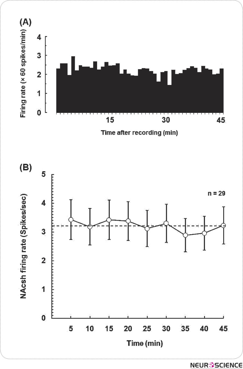
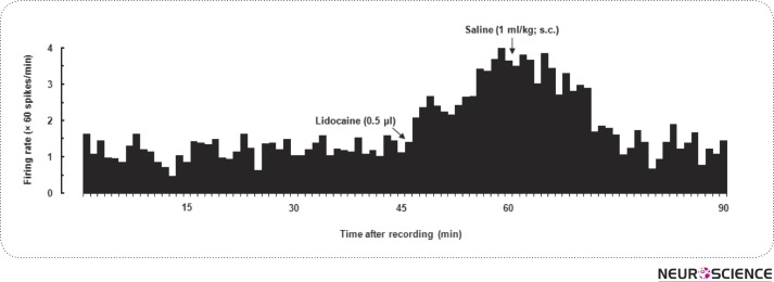
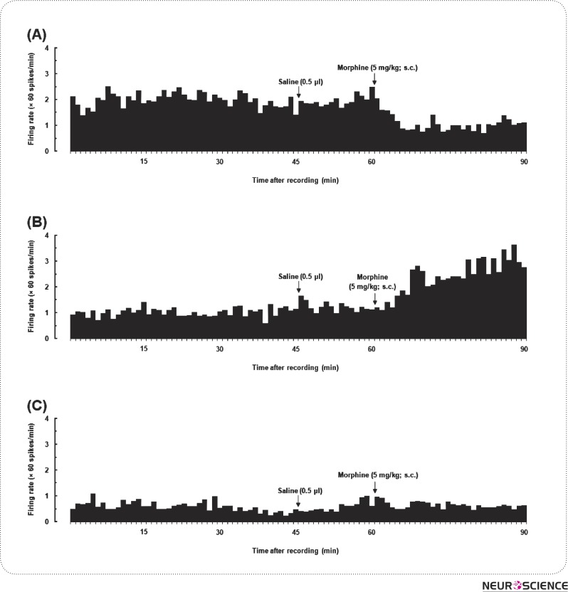
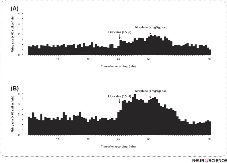
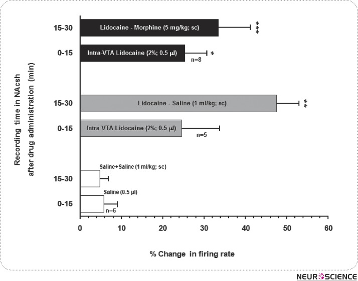
This study has examined the functional importance of nucleus accumbens (NAc)-ventral tegmental area (VTA) interactions. As it is known, this interaction is important in associative reward processes. Under urethane anesthesia, extracellular single unit recordings of the shell sub-region of the nucleus accumbens (NAcSh) neurons were employed to determine the functional contributions of the VTA to neuronal activity across NAcSh in rats. The baseline firing rate of NAcSh neurons varied between 0.42 and 11.44 spikes/sec and the average frequency of spontaneous activity over 45-minute period was 3.21±0.6 spikes/sec. The majority of NAcSh neurons responded excitatory in the first and second 15-min time blocks subsequent to the inactivation of VTA. In the next set of experiments, eight experimental rats received morphine (5 mg/kg; sc). Three patterns of neuronal activity were found. Among the recorded neurons only three had an increase followed by morphine administration. Whereas the other three neurons were attenuated following morphine administration; and there were no changes in the firing rates of the two neurons left. Finally, unilateral reversible inactivation of VTA attenuated the firing activity of the majority of ipsilateral NAcSh neuron in response to morphine, except for a single cell. These results suggest that transient inactivation of VTA reduces the ability of neurons in the NAcsh to respond to systemic morphine, and that NAcSh neuron activity depends on basal firing rate of VTA inputs.
Keywords: Morphine; Nucleus Accumbens; Rat; Reversible Inactivation; Single Unit Recording; Ventral Tegmental Area.
Figures
References
-
- Bunney, E.B. , Appel, S.B. , & Brodie, M.S. (2001). Electrophysiological effects of cocaethylene, cocaine, and ethanol on dopaminergic neurons of the ventral tegmental area. Journal of Pharmacology and Experimental Therapeutics, 297(2), 696–703 - PubMed
-
- Chu, N. , Xia, W. , Yu, P. , Hu, L. , Zhang, R. , & Cui, C. (2008). PRECLINICAL STUDY: Chronic morphine-induced neuronal morphological changes in the ventral tegmental area in rats are reversed by electroacupuncture treatment. Addiction biology, 13(1), 47–51 - PubMed
-
- Esmaeili, M.H. , Kermani, M. , Parvishan, A. , & Haghparast, A. (2012). Role of D1/D2 dopamine receptors in the CA1 region of the rat hippocampus in the rewarding effects of morphine administered into the ventral tegmental area. Behavioural brain research, 231(1), 111–5 - PubMed
-
- Gholami, A. , Zarrindast, M.R. , Sahraei, H. , & Haerri-Rohani, A. (2003). Nitric oxide within the ventral tegmental area is involved in mediating morphine reward. European journal of pharmacology, 458(1-2), 119–128 - PubMed
LinkOut - more resources
Full Text Sources