

No Correlation of Inflammation With Colonization of Helicobacter pylori in the Stomach of Mice Fed High-salt Diet
- PMID: 25337583
- PMCID: PMC4204169
- DOI: 10.15430/JCP.2014.19.2.144
No Correlation of Inflammation With Colonization of Helicobacter pylori in the Stomach of Mice Fed High-salt Diet
Abstract
Background: Previous studies on Helicobacter pylori infection in mice have contributed to better understanding of the pathogenesis of chronic gastritis and gastric carcinoma. The aim of this study was to evaluate H. pylori colonization and subsequent inflammatory responses in the stomachs of C57BL/6 mice depending on inoculation number and the presence of high-salt diet.
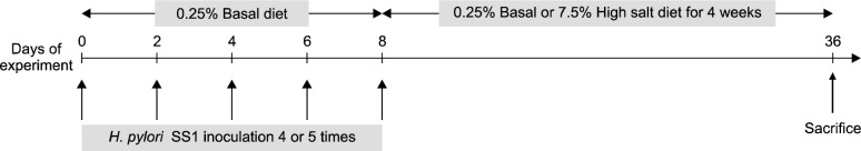
Methods: Eighty-four female mice with 4 weeks age were used in this study. The infected mice were gavaged with H. pylori strain Sydney-1 (SS1), and the uninfected mice were dosed with vehicle. In each of these groups, half of the mice were fed ona basal diet (0.25% salt) and the other half were fed on a high-salt diet (7.5% salt). The infected mice were inoculated 4 or 5 times, and infection status and degree of inflammation were checked by culture and histopathology, respectively, after 4 weeks. Gastric mucosal myeloperoxidase and tumor necrosis factor-alpha were measured by ELISA.
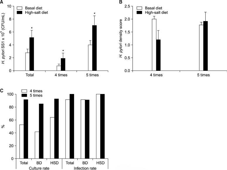
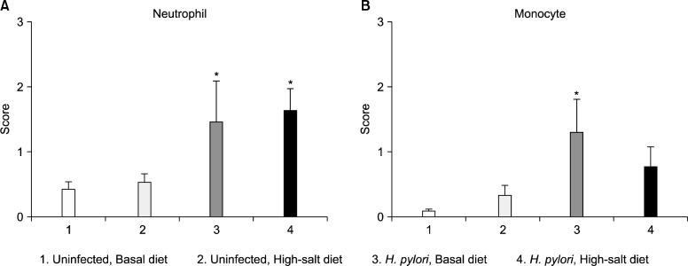
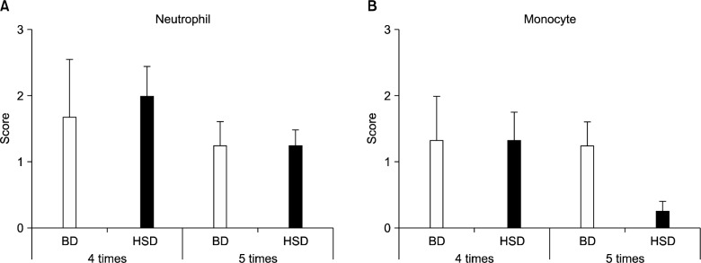
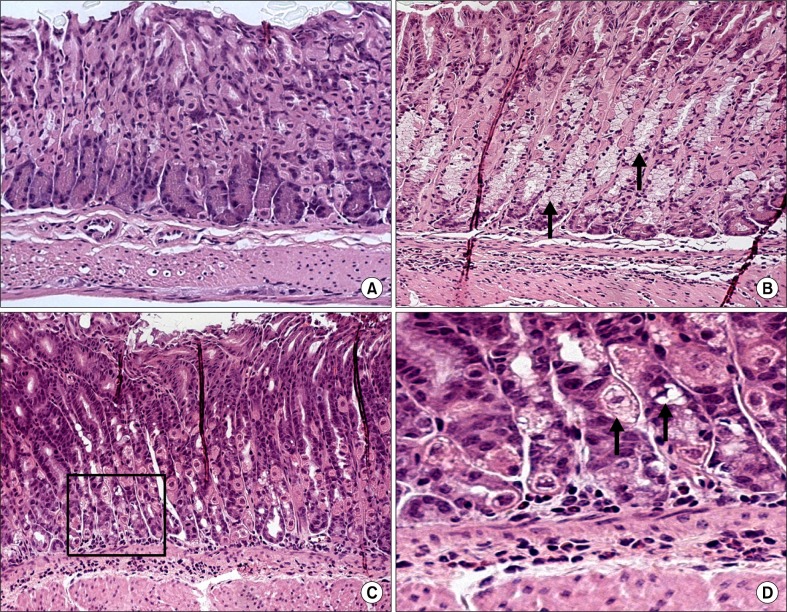
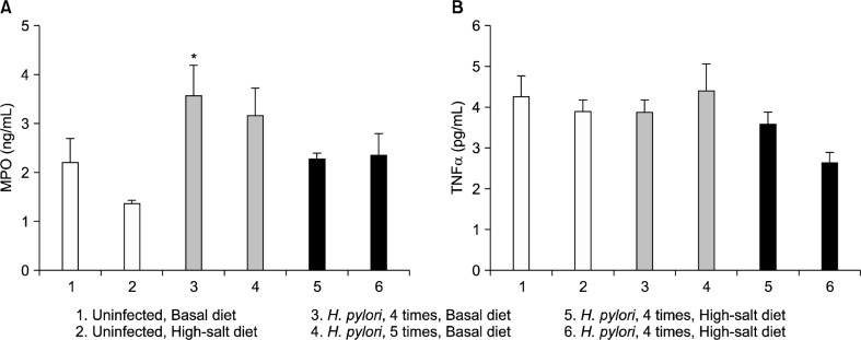
Results: The overall infection rate was 95.2%; the infection rate after 5 inoculations (100%) was greater than that after 4 inoculations (91.3%). However, no differences in the degree of inflammation were found between 2 groups. The bacterial density was also significantly increased in mice that were on the high-salt diet and had been inoculated 5 times, respectively. Mean neutrophil infiltration in the infected group was 1.7±0.6 (1, minimal; 2, mild; 3, moderate; 4, marked). However, the high-salt diet was not increase the inflammatory grade in the infected group. Gastric mucosal myeloperoxidase and tumor necrosis factor-alpha levels did not increased by the high-salt diet and increased the number of inoculation.
Conclusions: In spite of well colonization of H. pylori in the stomachs of C57BL/6 mice, the degree of subsequent inflammation was irrelevant to high-salt diet and frequent (5 times) inoculations.
Keywords: Helicobacter pylori; Inflammation; Salt.
Figures
References
-
- McColl KE. Clinical practice. Helicobacter pylori infection. N Engl J Med. 2010;362:1597–604. - PubMed
-
- Tsukamoto T, Toyoda T, Mizoshita T, Tatematsu M. Helicobacter pylori infection and gastric carcinogenesis in rodent models. Semin Immunopathol. 2013;35:177–90. - PubMed
-
- Lee A, Fox JG, Otto G, Murphy J. A small animal model of human Helicobacter pylori active chronic gastritis. Gastroenterology. 1990;99:1315–23. - PubMed
LinkOut - more resources
Full Text Sources
Other Literature Sources
Research Materials