Review
. 2014 Dec 5;13(12):5293-309.
doi: 10.1021/pr500880b. Epub 2014 Nov 4.

Isobaric labeling-based relative quantification in shotgun proteomics

Affiliations
Review

Isobaric labeling-based relative quantification in shotgun proteomics

Navin Rauniyar et al. J Proteome Res. .

Abstract

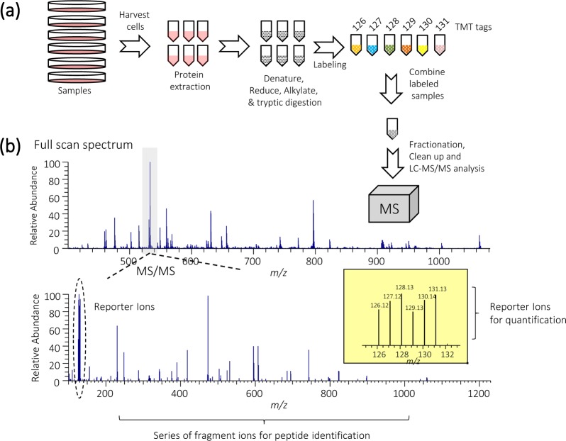
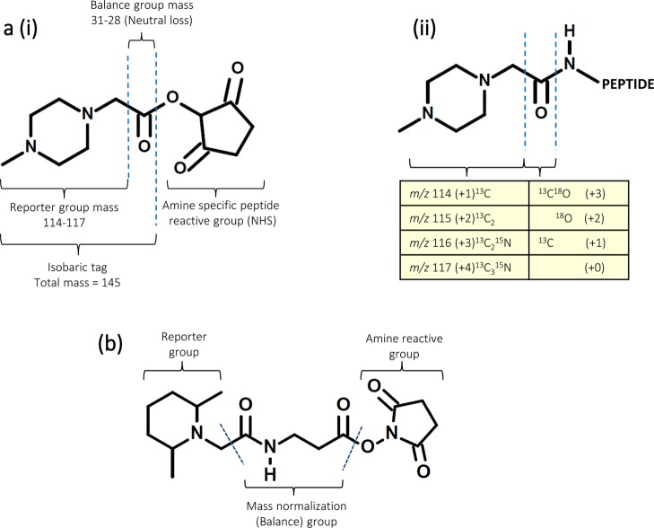
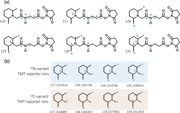
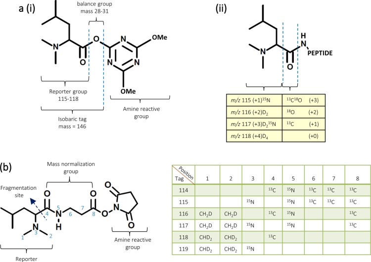
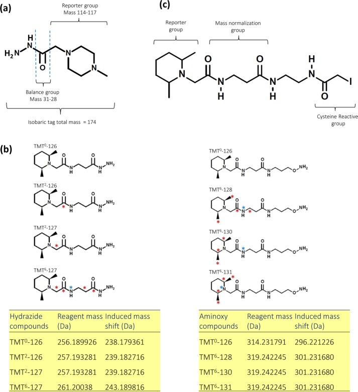
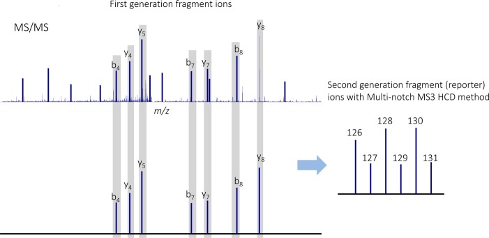
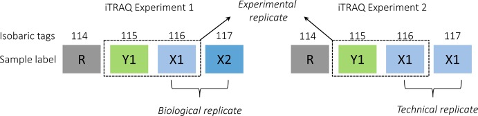
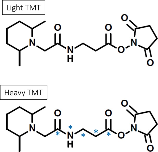
Mass spectrometry plays a key role in relative quantitative comparisons of proteins in order to understand their functional role in biological systems upon perturbation. In this review, we review studies that examine different aspects of isobaric labeling-based relative quantification for shotgun proteomic analysis. In particular, we focus on different types of isobaric reagents and their reaction chemistry (e.g., amine-, carbonyl-, and sulfhydryl-reactive). Various factors, such as ratio compression, reporter ion dynamic range, and others, cause an underestimation of changes in relative abundance of proteins across samples, undermining the ability of the isobaric labeling approach to be truly quantitative. These factors that affect quantification and the suggested combinations of experimental design and optimal data acquisition methods to increase the precision and accuracy of the measurements will be discussed. Finally, the extended application of isobaric labeling-based approach in hyperplexing strategy, targeted quantification, and phosphopeptide analysis are also examined.

Keywords: TMT; iTRAQ; isobaric labeling; isobaric tags; isobaric tags for relative and absolute quantification; mass spectrometry; quantitative proteomics; tandem mass tags.


Figures

Figure 1
Figure 1
(a) General workflow of an isobaric labeling experiment. The protocol involves extraction of proteins from cells or tissues followed by reduction, alkylation, and digestion. In the case of TMT 6-plex, up to six samples can be labeled with the six isobaric tags of the reagent. Resulting peptides are pooled at equal concentrations before fractionation and clean up. The TMT-labeled samples are analyzed by LC–MS/MS. (b) In an MS1 scan, same-sequence peptides from the different samples appear as a single unresolved additive precursor ion. Following fragmentation of the precursor ion during MS/MS, the six reporter ions appear as distinct masses between m/z 126–131, and the remainder of the sequence-informative b- and y-ions remains as additive isobaric signals. The reporter ion intensity indicates the relative amount of peptide in the mixture that was labeled with the corresponding reagent.
Figure 2
Figure 2
(a) (i) Chemical structure of iTRAQ 4-plex reagent. The complete molecule consists of a reporter group (based on N-methylpiperazine), a mass balance group (carbonyl), and a peptide-reactive group (NHS ester). The overall mass of the reporter and balance components of the molecule are kept constant using differential isotopic enrichment with 13C, 15N, and 18O atoms. The reporter group ranges in mass from m/z 114–117, whereas the balance group ranges in mass from 28 to 31 Da, such that the combined mass remains constant (145 Da) for each of the four reagents of the iTRAQ 4-plex set. (ii) The tag reacts with peptide N-terminus or ε-amino group of lysine to form an amide linkage that fragments in a similar fashion to that of backbone peptide bonds when subjected to CID. Following fragmentation of the tag amide bond, the balance (carbonyl) moiety is lost as neutral loss, whereas charge is retained by the reporter group. The number in parentheses in the table indicates the number of enriched centers in each section of the molecule. (b) Chemical structure of a generic TMT reagent showing the three functional groups: an amine-reactive group that labels the N-terminus and ε-amine group of lysine in peptides, a mass normalization (balance) group that balances mass differences from individual reporter ions to ensure the same overall mass of the reagents, and a reporter group that provides the abundance of a peptide upon MS/MS in individual samples being mixed. The blue dashed lines indicate a cleavable linker that enables the release of the reporter ion from the whole tag upon MS/MS. The TMT reagent family consists of TMTzero, TMTduplex, TMT 6-plex, and TMT 10-plex sets, and each of them is based on the same chemical structure.
Figure 3
Figure 3
(a) Chemical structure of TMT 6-plex reagents with 13C and 15N heavy isotope positions (blue asterisks). The tags are isobaric, with a different distribution of isotopes between the reporter and mass normalization (balance) groups. (b) The substitution of 15N for 13C to generate new reporter ions that are 6.32 mDa lighter than the original forms used in TMT 6-plex. The TMT 6-plex reagents in combination with four isotope variants of the tag with 6.32 mDa mass differences were used to generate TMT 10-plex reagent.
Figure 4
Figure 4
(a) (i) General structure of dimethyl leucine isobaric (DiLeu) mass tag. Reporter ions range from m/z 115–118. (ii) Illustration of formation of new peptide bond at N-terminus or ε-amino group of the lysine side chain and isotope combination of isobaric tags (b) Chemical structure of DiART isobaric reagents. Positions containing heavy stable isotopes are illustrated as numbers in the structure, and the table lists the elemental composition of the corresponding numbers. During MS/MS, the DiART-tagged peptides yield reporter ions ranging from m/z 114 to 119.
Figure 5
Figure 5
(a) General structure of iTRAQ hydrazide (iTRAQH) for relative quantitative analysis of carbonylation sites in proteins. (b) Chemical structure of the carbonyl-reactive glyco-TMT compounds. (Left) Hydrazide reagents; (right) aminoxy reagents. Red asterisks indicate 13C, and blue asterisks, 15N. The table below the compound structures shows isotope codes of the hydrazide- and aminoxy-functionalized glyco-TMT compounds. The carbonyl-reactive tags can be used to quantify a broad range of biologically important molecules including carbohydrates, steroids, or oxidized proteins. (c) Chemical structure of the cysteine-reactive Thermo Scientific iodoTMTzero isobaric mass tag. The iodoTMT reagents are iodoacetyl-activated isobaric mass tags for covalent, irreversible labeling of sulfhydryl (-SH) groups. IodoTMT 6-plex enable measurement of protein and peptide cysteine modifications (S-nitrosylation, oxidation, and disulfide bridges) by multiplex quantitative mass spectrometry. The workflow (not shown in the image) involves derivatization of modified peptides or proteins with the reagent, enrichment of TMT tagged peptide using anti-TMT antibody, and their subsequent elution. The eluent is analyzed by LC–MS/MS to determine the sites of modification and to measure their relative abundance across samples.
Figure 6
Figure 6
Multinotch MS3 involves selecting and co-isolating multiple MS/MS product ion and fragmenting them to generate a plurality of second-generation fragment ion species including released isobaric tags., The method increases the sensitivity and quantitative accuracy achieved by isobaric labeling-based quantification approach.
Figure 7
Figure 7
An example defining the relation among technical, experimental, and biological replicates in isobaric labeling (iTRAQ 4-plex in this example) experiments. A biological replicate has two distinct biological samples (X1 and X2) from the same condition in an iTRAQ set, whereas a technical replicate has two identical samples (X1 and X1) from the same biological source in an iTRAQ set. An experimental replicate is the repetitive analysis of the setup to assess the variation of the identical sample in two different iTRAQ sets (Y1 and X1 in experiment 1 versus Y1 and X1 in experiment 2). R refers to a reference sample that can be an individual sample or a pooled sample and allows cross-set comparison.
Figure 8
Figure 8
Chemical structure of isotopic reagents, light TMT and heavy TMT, used for targeted quantification. The light reagent has no heavy isotope incorporated, whereas the heavy reagent has five heavy isotopes incorporated (4 × 13C and 1 × 15N). Labeling with these reagents introduces mass differences into the peptides from different samples. In targeted experiments, quantification is obtained by structural b and/or y ions generated after collision-induced dissociation.

References

    1. Wu C. C.; MacCoss M. J.; Howell K. E.; Matthews D. E.; Yates J. R. III Metabolic labeling of mammalian organisms with stable isotopes for quantitative proteomic analysis. Anal. Chem. 2004, 76, 4951–4959. - PubMed
    1. Ong S. E.; Blagoev B.; Kratchmarova I.; Kristensen D. B.; Steen H.; Pandey A.; Mann M. Stable isotope labeling by amino acids in cell culture, SILAC, as a simple and accurate approach to expression proteomics. Mol. Cell. Proteomics 2002, 1, 376–386. - PubMed
    1. Rose C. M.; Merrill A. E.; Bailey D. J.; Hebert A. S.; Westphall M. S.; Coon J. J. Neutron encoded labeling for peptide identification. Anal. Chem. 2013, 85, 5129–5137. - PMC - PubMed
    1. Gygi S. P.; Rist B.; Gerber S. A.; Turecek F.; Gelb M. H.; Aebersold R. Quantitative analysis of complex protein mixtures using isotope-coded affinity tags. Nat. Biotechnol. 1999, 17, 994–999. - PubMed
    1. Hsu J. L.; Huang S. Y.; Chow N. H.; Chen S. H. Stable-isotope dimethyl labeling for quantitative proteomics. Anal. Chem. 2003, 75, 6843–6852. - PubMed

Publication types