

Microcephaly-chorioretinopathy syndrome, autosomal recessive form. A case report
- PMID: 25337662
- PMCID: PMC10876356
- DOI: 10.1590/1516-3180.2013.7930003
Microcephaly-chorioretinopathy syndrome, autosomal recessive form. A case report
Abstract
Context: The autosomal recessive form of microcephaly-chorioretinopathy syndrome is a rare genetic condition that is considered to be an important differential diagnosis with congenital toxoplasmosis.
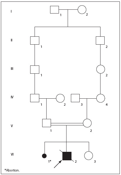
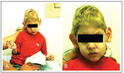
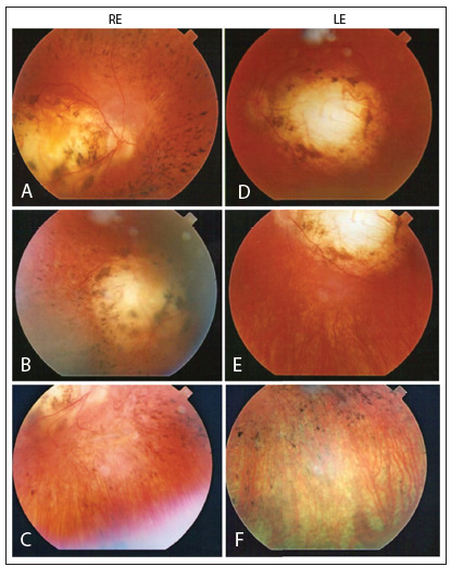
Case report: Our patient was a seven-year-old white boy who was initially diagnosed with congenital toxoplasmosis. However, his serological tests for congenital infections, including toxoplasmosis, were negative. He was the first child of young, healthy and consanguineous parents (fourth-degree relatives). The parents had normal head circumferences and intelligence. The patient presented microcephaly and specific abnormalities of the retina, with multiple diffuse oval areas of pigmentation and patches of chorioretinal atrophy associated with diffuse pigmentation of the fundus. Ophthalmological evaluations on the parents were normal. A computed tomography scan of the child's head showed slight dilation of lateral ventricles and basal cisterns without evidence of calcifications. We did not find any lymphedema in his hands and feet. He had postnatal growth retardation, severe mental retardation and cerebral palsy.
Conclusions: The finding of chorioretinal lesions in a child with microcephaly should raise suspicions of the autosomal recessive form of microcephaly-chorioretinopathy syndrome, especially in cases with an atypical pattern of eye fundus and consanguinity. A specific diagnosis is essential for an appropriate clinical evaluation and for genetic counseling for the patients and their families.
CONTEXTO:: A forma autossômica recessiva da síndrome de microcefalia-coriorretinopatia é condição genética rara, considerada um importante diagnóstico diferencial com toxoplasmose congênita.
RELATO DO CASO:: O paciente era um menino branco de sete anos de idade, inicialmente diagnosticado com toxoplasmose congênita. No entanto, suas sorologias para infecções congênitas, incluindo a toxoplasmose, eram negativas. Ele foi o primeiro filho de pais jovens, hígidos e consanguíneos (parentes de quarto grau). Os pais apresentavam perímetro cefálico e inteligência normais. O paciente apresentava microcefalia e anormalidades específicas da retina com áreas ovais de pigmentação múltiplas e difusas, além de manchas de atrofia coriorretiniana associadas à pigmentação difusa do fundo de olho. A avaliação oftalmológica dos pais foi normal. A tomografia computadorizada de crânio da criança mostrou discreta dilatação dos ventrículos laterais e cisternas basais, sem evidência de calcificações. Nós não verificamos a presença de linfedema em suas mãos e pés. Ele possuía retardo do crescimento pós-natal, deficiência mental grave e paralisia cerebral.
CONCLUSÃO:: O achado de lesões coriorretinianas em uma criança com microcefalia deve aumentar a suspeita da forma autossômica recessiva da síndrome de microcefalia-coriorretinopatia, principalmente em casos com padrão atípico de fundo de olho e consanguinidade. O diagnóstico preciso é essencial para correta avaliação clínica e aconselhamento genético dos pacientes e suas famílias.
Conflict of interest statement
Figures
Similar articles
-
An autosomal recessive form of spastic cerebral palsy (CP) with microcephaly and mental retardation.Am J Med Genet A. 2006 Jul 15;140(14):1504-10. doi: 10.1002/ajmg.a.31288. Am J Med Genet A. 2006. PMID: 16761294 Free PMC article.
-
The syndrome of retinal pigmentary degeneration, microcephaly, and severe mental retardation (Mirhosseini-Holmes-Walton syndrome): report of two patients.Am J Med Genet. 1985 Oct;22(2):223-8. doi: 10.1002/ajmg.1320220202. Am J Med Genet. 1985. PMID: 4050854
-
Autosomal recessive microcephaly associated with chorioretinopathy.Hum Genet. 1977 Apr 15;36(2):243-7. doi: 10.1007/BF00273265. Hum Genet. 1977. PMID: 870417
-
An Italian family affected by autosomal dominant microcephaly with chorioretinal degeneration.J Pediatr Ophthalmol Strabismus. 2002 Sep-Oct;39(5):288-92. doi: 10.3928/0191-3913-20020901-09. J Pediatr Ophthalmol Strabismus. 2002. PMID: 12353901 Review.
-
Filippi syndrome: a new case with skeletal abnormalities.J Med Genet. 1995 Aug;32(8):659-61. doi: 10.1136/jmg.32.8.659. J Med Genet. 1995. PMID: 7473664 Free PMC article. Review.
References
-
- Petersen E. Toxoplasmosis. Semin Fetal Neonatal Med. 2007;12(3):214–223. - PubMed
-
- McKusick VA, Stauffer M, Knox L, Clark DB. Chorioretinopathy with hereditary microcephaly. Arch Ophthalmol. 1966;75(5):597–600. - PubMed
-
- Schmidt B, Jaeger W, Neubauer H. Ein Mikrozephalie-Syndrom mit atypischer tapetoretinaler degeneration bei 3 Geschwistern [A microcephalic syndrome with atypical tapetoretinal degeneration in 3 siblings] Klin Monbl Augenheilkd. 1967;150(2):188–196. - PubMed
-
- Cantú JM, Rojas JA, García-Cruz D, et al. Autosomal recessive microcephaly associated with chorioretinopathy. Hum Genet. 1977;36(2):243–247. - PubMed
-
- Abdel-Salam GM, Czeizel AE, Vogt G, Imre L. Microcephaly with chorioretinal dysplasia: characteristic facial features. Am J Med Genet. 2000;95(5):513–515. - PubMed
Publication types
MeSH terms
LinkOut - more resources
Full Text Sources
Other Literature Sources
