. 2014 Nov 26;136(47):16544-56.
doi: 10.1021/ja507847t. Epub 2014 Nov 13.

A set of computationally designed orthogonal antiparallel homodimers that expands the synthetic coiled-coil toolkit

Affiliations

A set of computationally designed orthogonal antiparallel homodimers that expands the synthetic coiled-coil toolkit

Christopher Negron et al. J Am Chem Soc. .

Abstract

Molecular engineering of protein assemblies, including the fabrication of nanostructures and synthetic signaling pathways, relies on the availability of modular parts that can be combined to give different structures and functions. Currently, a limited number of well-characterized protein interaction components are available. Coiled-coil interaction modules have been demonstrated to be useful for biomolecular design, and many parallel homodimers and heterodimers are available in the coiled-coil toolkit. In this work, we sought to design a set of orthogonal antiparallel homodimeric coiled coils using a computational approach. There are very few antiparallel homodimers described in the literature, and none have been measured for cross-reactivity. We tested the ability of the distance-dependent statistical potential DFIRE to predict orientation preferences for coiled-coil dimers of known structure. The DFIRE model was then combined with the CLASSY multistate protein design framework to engineer sets of three orthogonal antiparallel homodimeric coiled coils. Experimental measurements confirmed the successful design of three peptides that preferentially formed antiparallel homodimers that, furthermore, did not interact with one additional previously reported antiparallel homodimer. Two designed peptides that formed higher-order structures suggest how future design protocols could be improved. The successful designs represent a significant expansion of the existing protein-interaction toolbox for molecular engineers.


Figures

Figure 1
Figure 1
Predicting coiled-coil orientation preference and testing cluster-expanded DFIRE*. (A) EAP and EP are the antiparallel (AP) and parallel (P) DFIRE* energies for each orientation test set coiled coil. Antiparallel or parallel coiled coils (according to PDB structure) are plotted with red crosses or black diamonds, respectively. The line at EAPEP = 0.18 AU gives optimal separation of parallel and antiparallel examples. Min_gap was used to remove examples with small DFIRE* orientation preferences (see text); shading indicates increasing min_gap from the line of optimal separation. (B) The fraction of antiparallel sequences predicted correctly vs the fraction of parallel sequences predicted correctly, as the cutoff value for EAPEP was changed, is plotted for DFIRE* and the CE model of DFIRE*. Curves for data sets with different values of min_gap are shown for the CE model of DFIRE*. (C, D) DFIRE* energies vs the CE model of DFIRE* energies for randomly generated dimer-like test structures in the antiparallel (C) and parallel (D) states.
Figure 2
Figure 2
Computational design of orthogonal antiparallel homodimers. (A, B) Diagram of target and off-target states included in two design calculations. Colors represent distinct sequences, and colored circles indicate the N-terminus of each helix. An energetic constraint, Δ, was enforced between the energy of each target antiparallel homodimer state (E1, E2, E3) and every off-target state that peptide could participate in (examples shown with gray dashed lines). The sequence space used for each design is indicated. Different numbers of off-target states were included for sequence space 1 (A) vs sequence space 2 (B). (C, D) The total energy ET = E1 + E2 + E3 vs Δ is plotted for sequence space 1 (C) and sequence space 2 (D). Each value of Δ led to a set of optimized sequences, and the gray squares mark the solutions chosen for experimental testing.
Figure 3
Figure 3
Designed peptides APH2, APH3, and APH4 adopt an antiparallel helix orientation. (A) Schematic of the assay. Arrows indicate helix direction from N to C terminus. The wavy line indicates two amino acids added to the designed sequence to change peptide retention times (APH2 = YY, APH3 = QW, APH4 = YY). S represents the sulfur atom in cysteine residue(s). (B, C, D) HPLC chromatograms show the results for the disulfide-exchange reactions upon mixing equimolar amounts of N-terminal and C-terminal cysteine variants of each design sequence (20 μM each). The reactions were quenched at 0 min (red), 15 min (black), or 5 h (blue). Peaks are labeled according to the scheme shown in panel A, with G indicating a glutathione adduct.
Figure 4
Figure 4
Designed peptides APH2, APH3 and APH4 do not form heterodimers. (A) Cartoon showing four cysteine-containing peptides, two for each of two designs, which were included in the disulfide-exchange cross-reactivity assay. (B, C, D) HPLC traces for all pairwise mixtures of designed peptides after equilibration for 15 min. The blue and red traces are for reactions with equimolar amounts of N- and C-terminal cysteine variants of a single designed peptide (20 μM each). The black trace is for a reaction with equimolar amounts of all four peptides in panel A (20 μM each). (B) APH2 + APH3, (C) APH2 + APH4, (D) APH3 + APH4.
Figure 5
Figure 5
Designed peptides APH2, APH3, and APH4 do not heterodimerize with APH. (A, B, C) HPLC traces for all pairwise combinations of APH with the designed coiled coils, with experimental conditions as for Figure 4. The blue and red traces are for equimolar mixtures of N- and C-terminal cysteine variants of APH (blue) or APH2, APH3 or APH4 (red) (20 μM each). The black trace is for a mixture of four peptides, APH and the indicated design, each modified at the N- or C-terminus with a cysteine residue (20 μM each). (A) APH + APH2, (B) APH + APH3, (C) APH + APH4.
Figure 6
Figure 6
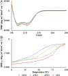
Circular dichroism spectra and thermal denaturation curves. (A) CD spectra and (B) thermal denaturation curves measured at 25 °C in PBS with 1 mM DTT. APH (red), APH2 (blue), APH3 (green) and APH4 (orange).
Figure 7
Figure 7
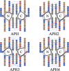
Helical-wheel diagrams of APH, APH2, APH3, and APH4 as antiparallel homodimers. Positively and negatively charged amino acids are shown in blue and red, respectively, with noncharged polar residues in orange and hydrophobic residues in gray. Potentially attractive salt bridges are shown as dashed lines. Sequences start at an f position and end at an e position. Diagrams were generated using DrawCoil 1.0, http://www.grigoryanlab.org/drawcoil.

Similar articles

Cited by

References

    1. Purnick P. E.; Weiss R. Nat. Rev. Mol. Cell Biol. 2009, 10, 410–422. - PubMed
    1. Thompson K. E.; Bashor C. J.; Lim W. A.; Keating A. E. ACS Synth. Biol. 2012, 1, 118–129. - PMC - PubMed
    1. Fletcher J. M.; Boyle A. L.; Bruning M.; Bartlett G. J.; Vincent T. L.; Zacci N. R.; Armstrong C. T.; Bromley E. H. C.; Booth P. J.; Brady R. L.; Thomson A. R.; Woolfson D. N. ACS Synth. Biol. 2012, 1, 240–250. - PubMed
    1. Wolf E.; Kim P. S.; Berger B. Protein Sci. 1997, 6, 1179–1189. - PMC - PubMed
    1. Rackham O. J.; Madera M.; Armstrong C. T.; Vincent T. L.; Woolfson D. N.; Gough J. J. Mol. Biol. 2010, 403, 480–493. - PubMed

Publication types