. 2014 Oct 22;9(10):e110916.
doi: 10.1371/journal.pone.0110916. eCollection 2014.

Characteristic and functional analysis of a newly established porcine small intestinal epithelial cell line

Affiliations

Characteristic and functional analysis of a newly established porcine small intestinal epithelial cell line

Jing Wang et al. PLoS One. .

Abstract

The mucosal surface of intestine is continuously exposed to both potential pathogens and beneficial commensal microorganisms. Recent findings suggest that intestinal epithelial cells, which once considered as a simple physical barrier, are a crucial cell lineage necessary for maintaining intestinal immune homeostasis. Therefore, establishing a stable and reliable intestinal epithelial cell line for future research on the mucosal immune system is necessary. In the present study, we established a porcine intestinal epithelial cell line (ZYM-SIEC02) by introducing the human telomerase reverse transcriptase (hTERT) gene into small intestinal epithelial cells derived from a neonatal, unsuckled piglet. Morphological analysis revealed a homogeneous cobblestone-like morphology of the epithelial cell sheets. Ultrastructural indicated the presence of microvilli, tight junctions, and a glandular configuration typical of the small intestine. Furthermore, ZYM-SIEC02 cells expressed epithelial cell-specific markers including cytokeratin 18, pan-cytokeratin, sucrase-isomaltase, E-cadherin and ZO-1. Immortalized ZYM-SIEC02 cells remained diploid and were not transformed. In addition, we also examined the host cell response to Salmonella and LPS and verified the enhanced expression of mRNAs encoding IL-8 and TNF-α by infection with Salmonella enterica serovars Typhimurium (S. Typhimurium). Results showed that IL-8 protein expression were upregulated following Salmonella invasion. TLR4, TLR6 and IL-6 mRNA expression were upregulated following stimulation with LPS, ZYM-SIEC02 cells were hyporeponsive to LPS with respect to IL-8 mRNA expression and secretion. TNFα mRNA levels were significantly decreased after LPS stimulation and TNF-α secretion were not detected challenged with S. Typhimurium neither nor LPS. Taken together, these findings demonstrate that ZYM-SIEC02 cells retained the morphological and functional characteristics typical of primary swine intestinal epithelial cells and thus provide a relevant in vitro model system for future studies on porcine small intestinal pathogen-host cell interactions.


Conflict of interest statement

Competing Interests: The authors have declared that no competing interests exist.

Figures

Figure 1
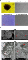
Figure 1. Morphological features of pSIECs and ZYM-SIEC02 cells.
A. Cellular morphology of pSIECs at 7 days in culture (100×) and at passage 5 (100×); a drug-resistant cell clone (100×) and ZYM-SIEC02 cells at passage 90 (100×).No obvious morphological differences were observed between ZYM-SIEC02 cells and primary SIECs. B. Electron micrographs of monolayer cultures of ZYM-SIEC02 cells indicate microvilli (MV) in 3 dimensions andin monolayer culture; tight junctions (TJ) and a small intestine glandular configuration (SIGC) (Red arrow).
Figure 2
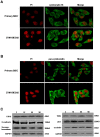
Figure 2. Immunostaining and western blot analysis of ZYM-SIEC02 cell cultures.
A. pSIECs and ZYM-SIEC02 cells were stained with cytokeratin 18 antibodies (green) and propidium iodide (red). B. pSIECs and ZYM-SIEC02 cells were stained with pan-cytokeratin antibodies (green) and propidium iodide (red). C. expression of cytokeratin 18, E-cadherin, SI, villin, ZO-1 and occludin were determined by western blot analysis. Cytokeratin 18, pan-cytokeratin, E-cadherin, SI, villin, ZO-1 and occludin were expressed in both pSIECs (at passage 3) and ZYM-SIEC02 cells (at passages 15, 55, and 90).
Figure 3
Figure 3. Growth curves, apoptosis and cell cycle analysis of SIECs before and after hTERT transfection.
A. Growth of pSIECs and ZYM-SIEC02 cells. Data are represented as the mean ± SD of 3 independent experiments. B. Apoptosis analysis of control pSIECs and ZYM-SIEC02 cells, The percentage of apoptosis in pSIECs and ZYM-SIEC02 cells under basal growth conditions was 26.9% and 11.4% (Q2+Q4), respectively. C. Cell cycle distributions of control SIECs and ZYM-SIEC02 cells, the percentages of pSIECs and ZYM-SIEC02 cells in S phase were 20.66% and 31.65%, respectively.
Figure 4
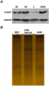
Figure 4. Detection of Telomerase activity.
A. Expression of hTERT protein was detected by western blot analysis. Expression of hTERT was readily detected in ZYM-SIEC02 cells at passage 50 and 90, but was barely detectable in pSIECs at passage 3. The A549 cell line was used as a positive control. B. Silver staining method was used to detect telomerase activity. The laddering patterns indicated that ZYM-SIEC02 cells display higher telomerase activity as compared to pSIECs and A549 cell line.
Figure 5
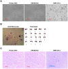
Figure 5. Tumorigenicity assay of ZYM-SIEC02 cells.
A. Representative images of pSIECs and ZYM-SIEC02 cells growing in soft agar. Neither cell line formed colonies after 2 weeks in culture, indicating that the cells did not exhibit anchorage-independent growth (100×). B. Karyotype analysis of ZYM-SIEC02 cells at passage 55, indicated that the cells contained 38 chromosomes, consistent with a diploid karyotype. C. Tumor formation assay. pSIECs and ZYM-SIEC02 cells were injected subcutaneously into nude mice. After one month, neither group of mice had developed tumors, in contrast, mice injected with EMF6 cells rapidly developed tumors within 8 days. Histological examination indicated morphologically normal tissue below the injection site of pSIECs (passage 3) and ZYM-SIEC02 cells (passage 55), but a dense cellular mass below the EMF6 cell injection site.
Figure 6
Figure 6. Optimization of culture medium for ZYM-SIEC02 cells.
A. ZYM-SIEC02 cells were cultured for 24 h with 2%, 5%, 10%, 15%, or 20% FBS. 5% FBS was found to be the optimal concentration. B. Addition of 5% FBS or ITS, and/or EGF to the cell medium individually, compared with DMEM/F12 alone; Addition of ITS or ITS/EGF enhanced cell growthsimilar to the addition of 5% FBS, while addition of EGF alone did not have a significant effect. C. ITS/EGF or ITS significantly enhanced cell growth with the use of cell culture medium containing 5% FBS. D. Addition of ITS, EGF, or EGF/ITS to DMEM/F12 had little effect on the proliferation rate compared to the addition to 5% FBS. * P□0.05 versus basal conditions.
Figure 7
Figure 7. Invasion efficiency of Samonlla Thpyimurium in the ZYM-SIEC02 cell line.
Confluent monolayers of ZYM-SIEC02 cells and pSIECs were infected with a MOI of 10∶1 (S. Typhimurium to host cells) for 1 h, and incubated in media containing gentamicin for an additional hour. Cells used were pSIECs (open bars) and ZYM-SIEC02 cells (filled bars). Data shown are normalized to the efficiency (relative percent of input bacteria) and representative of 5 independent experiments performed in triplicate.
Figure 8
Figure 8. Cytokine expression in ZYM-SIEC02 cells infected with Salmonella typhimurium and LPS.
Confluent ZYM-SIEC02 cell monolayers were infected for 4 h with S. Typhimurium or LPS. A. Relative mRNA expression of IL-8 in ZYM-SIEC02 cells were upregulated approximately 2.5-fold within 4 h of S. Typhimurium challenge relative to control, while the cells that stimulated with LPS demonstrated a significant reduction; B. TNF-α mRNA expression was a modest increased by S. Typhimurium stimulation, while the cells that stimulated with LPS demonstrated a significant reduction; C. TLR4 and TLR6 mRNA expression were upregulated following stimulation with LPS. D. IL-8 protein expression in ZYM-SIEC02 cells infected with Salmonella typhimurium and LPS. ZYM-SIEC02 cells were incubated with S. Typhimurium at a MOI of 100∶1 followed by an incubation in media containing gentamicin, and then cell culture supernatants were harvested after 4 h. ZYM-SIEC02 cells were stimulated with LPS, and cells were harvested after 4 h. IL-8 concentrations were determined by ELISA. The data shown are means±SEM of 3 independent experiments. * P<0.05;** P<0.01; *** P<0.001; ns, not significant.
Figure 9
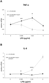
Figure 9. Inhibition of LPS-mediated response.
ZYM-SIEC02 cells were stimulated with the indicated dose of E.coli 055:B55 LPS in the presence or absence of polymyxin B sulfate(Sigma) for 3 hours. A. Polymyxin B increased TNF-α mRNA expression when the concentration of LPS was lower than 250 ng/ml. B. Polymyxin B prevented the increase in IL-6 mRNA expression. The data shown are means±SEM of 3 independent experiments. * P<0.05;** P<0.01; *** P<0.001.

Similar articles

Cited by

References

    1. Burch DG (1982) Tiamulin feed premix in the prevention and control of swine dysentery under farm conditions in the UK. Vet Rec 110: 244–246. - PubMed
    1. Schierack P, Nordhoff M, Pollmann M, Weyrauch KD, Amasheh S, et al. (2006) Characterization of a porcine intestinal epithelial cell line for in vitro studies of microbial pathogenesis in swine. Histochem Cell Biol 125: 293–305. - PubMed
    1. Sun Z, Huber VC, McCormick K, Kaushik RS, Boon AC, et al. (2012) Characterization of a porcine intestinal epithelial cell line for influenza virus production. J Gen Virol 93: 2008–2016. - PMC - PubMed
    1. Gibson-D'Ambrosio RE, Samuel M, D'Ambrosio SM (1986) A method for isolating large numbers of viable disaggregated cells from various human tissues for cell culture establishment. In Vitro Cell Dev Biol 22: 529–534. - PubMed
    1. Panja A (2000) A novel method for the establishment of a pure population of nontransformed human intestinal primary epithelial cell (HIPEC) lines in long term culture. Lab Invest 80: 1473–1475. - PubMed

Publication types

MeSH terms

LinkOut - more resources