

High-resolution mechanical imaging of glioblastoma by multifrequency magnetic resonance elastography
- PMID: 25338072
- PMCID: PMC4206430
- DOI: 10.1371/journal.pone.0110588
High-resolution mechanical imaging of glioblastoma by multifrequency magnetic resonance elastography
Abstract
Objective: To generate high-resolution maps of the viscoelastic properties of human brain parenchyma for presurgical quantitative assessment in glioblastoma (GB).
Methods: Twenty-two GB patients underwent routine presurgical work-up supplemented by additional multifrequency magnetic resonance elastography. Two three-dimensional viscoelastic parameter maps, magnitude |G*|, and phase angle φ of the complex shear modulus were reconstructed by inversion of full wave field data in 2-mm isotropic resolution at seven harmonic drive frequencies ranging from 30 to 60 Hz.
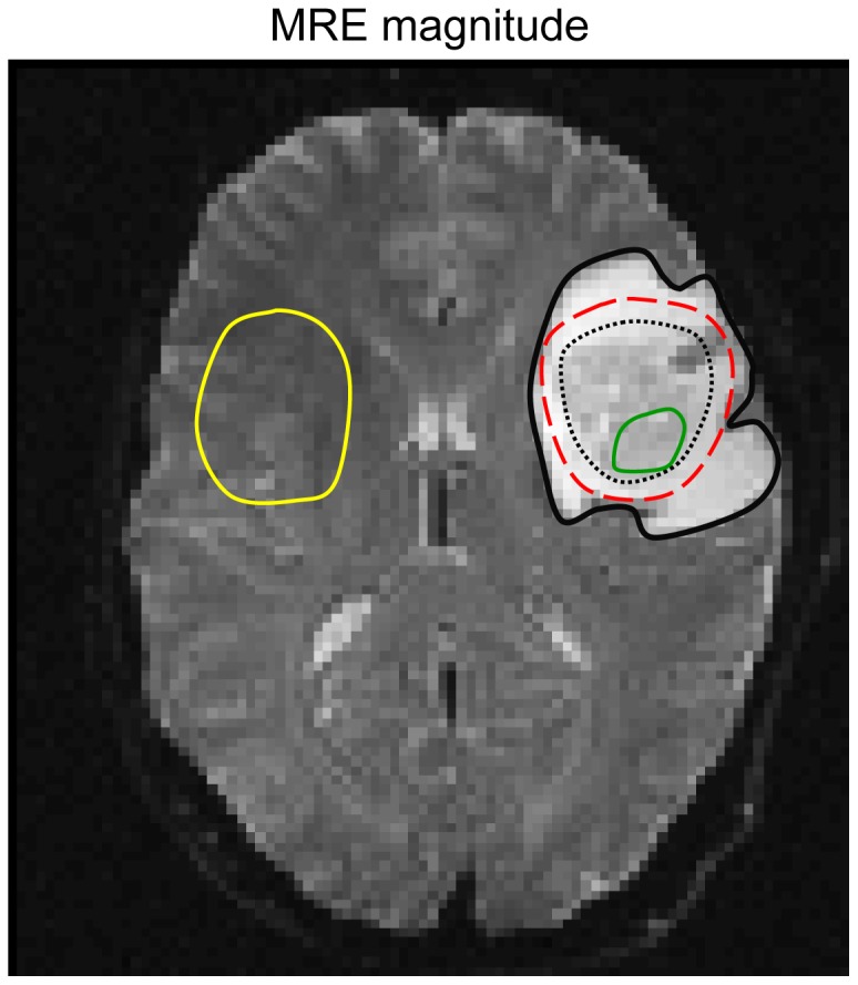
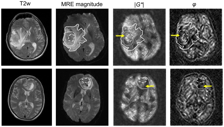
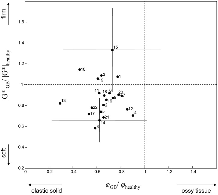
Results: Mechanical brain maps confirmed that GB are composed of stiff and soft compartments, resulting in high intratumor heterogeneity. GB could be easily differentiated from healthy reference tissue by their reduced viscous behavior quantified by φ (0.37±0.08 vs. 0.58±0.07). |G*|, which in solids more relates to the material's stiffness, was significantly reduced in GB with a mean value of 1.32±0.26 kPa compared to 1.54±0.27 kPa in healthy tissue (P = 0.001). However, some GB (5 of 22) showed increased stiffness.
Conclusion: GB are generally less viscous and softer than healthy brain parenchyma. Unrelated to the morphology-based contrast of standard magnetic resonance imaging, elastography provides an entirely new neuroradiological marker and contrast related to the biomechanical properties of tumors.
Conflict of interest statement
Figures
References
-
- Furnari FB, Fenton T, Bachoo RM, Mukasa A, Stommel JM, et al. (2007) Malignant astrocytic glioma: genetics, biology, and paths to treatment. Genes Dev 21: 2683–2710. - PubMed
-
- Stupp R, Mason WP, van den Bent MJ, Weller M, Fisher B, et al. (2005) Radiotherapy plus concomitant and adjuvant temozolomide for glioblastoma. N Engl J Med 352: 987–996. - PubMed
-
- CBTRUS Central Brain Tumor Registry of the United States: CBTRUS Statistical Report: Primary Brain and Central Nervous System Tumors Diagnosed in the United States in 2004–22008.
Publication types
MeSH terms
LinkOut - more resources
Full Text Sources
Other Literature Sources
Medical
