Review
. 2015 Feb;22(1):R1-31.
doi: 10.1530/ERC-14-0448. Epub 2014 Oct 22.

The new biology of estrogen-induced apoptosis applied to treat and prevent breast cancer

Affiliations
Review

The new biology of estrogen-induced apoptosis applied to treat and prevent breast cancer

V Craig Jordan. Endocr Relat Cancer. 2015 Feb.

Abstract

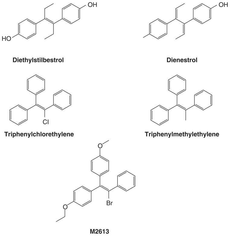
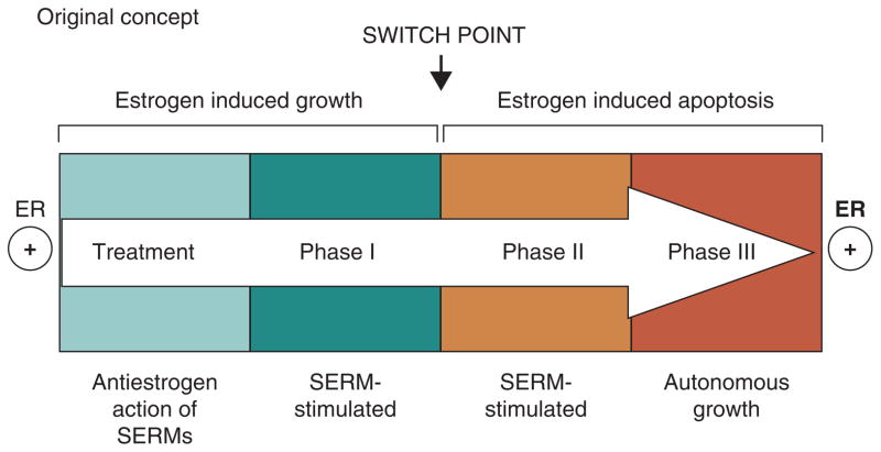
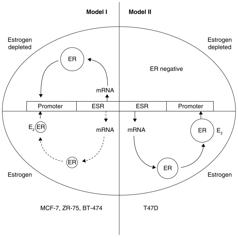
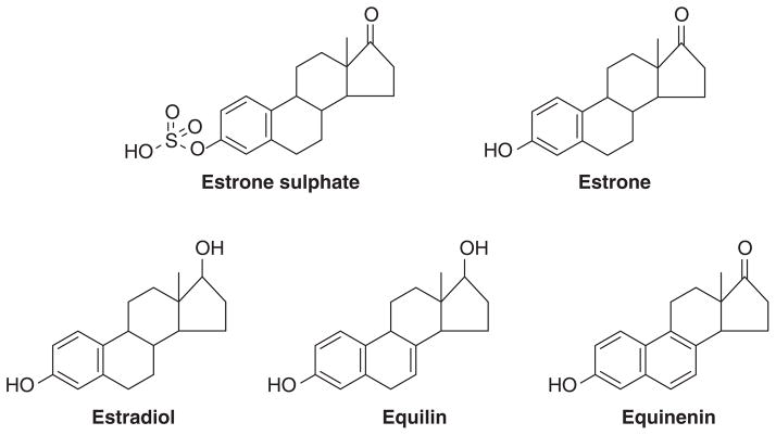
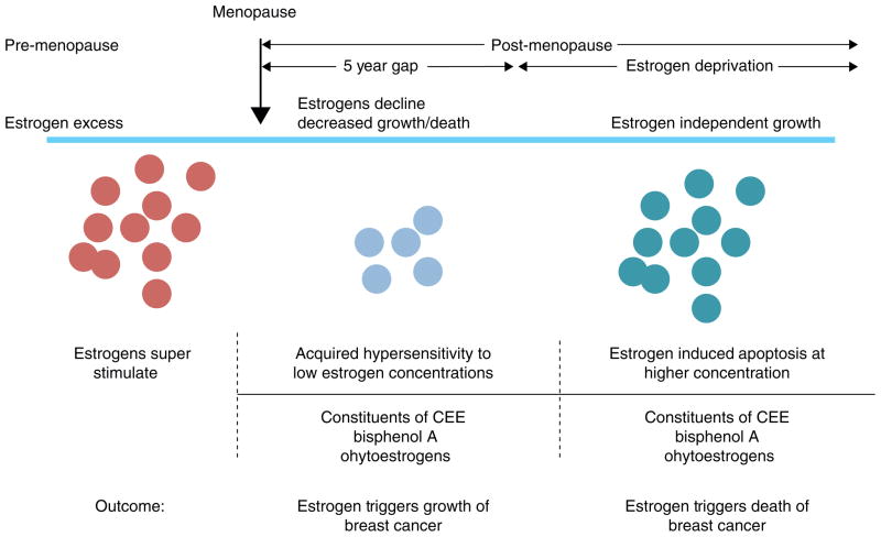
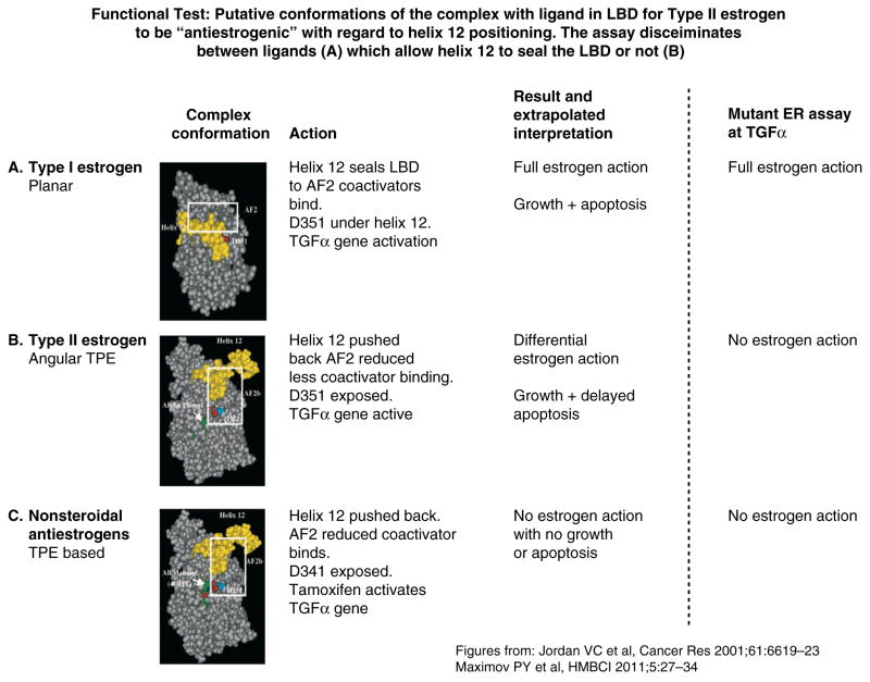
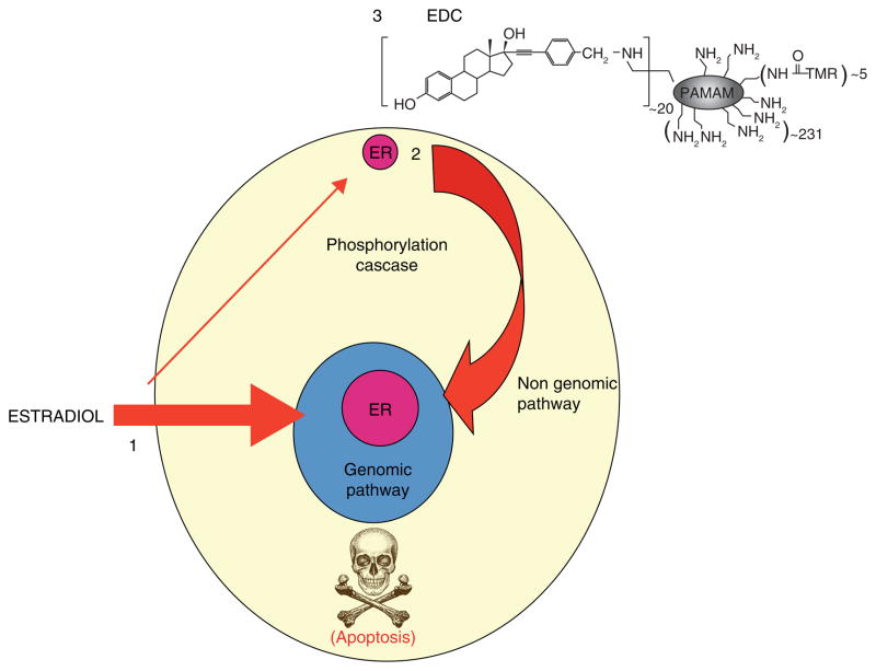
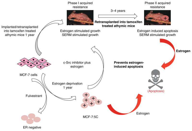
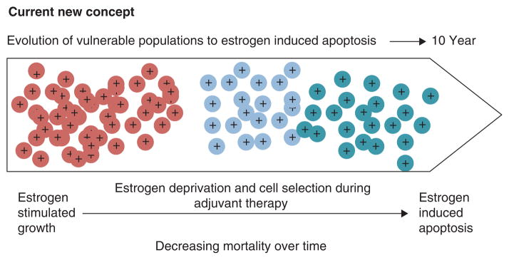
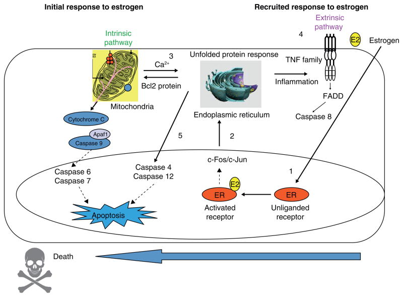
The successful use of high-dose synthetic estrogens to treat postmenopausal metastatic breast cancer is the first effective 'chemical therapy' proven in clinical trial to treat any cancer. This review documents the clinical use of estrogen for breast cancer treatment or estrogen replacement therapy (ERT) in postmenopausal hysterectomized women, which can either result in breast cancer cell growth or breast cancer regression. This has remained a paradox since the 1950s until the discovery of the new biology of estrogen-induced apoptosis at the end of the 20th century. The key to triggering apoptosis with estrogen is the selection of breast cancer cell populations that are resistant to long-term estrogen deprivation. However, estrogen-independent growth occurs through trial and error. At the cellular level, estrogen-induced apoptosis is dependent upon the presence of the estrogen receptor (ER), which can be blocked by nonsteroidal or steroidal antiestrogens. The shape of an estrogenic ligand programs the conformation of the ER complex, which, in turn, can modulate estrogen-induced apoptosis: class I planar estrogens (e.g., estradiol) trigger apoptosis after 24 h, whereas class II angular estrogens (e.g., bisphenol triphenylethylene) delay the process until after 72 h. This contrasts with paclitaxel, which causes G2 blockade with immediate apoptosis. The process is complete within 24 h. Estrogen-induced apoptosis is modulated by glucocorticoids and cSrc inhibitors, but the target mechanism for estrogen action is genomic and not through a nongenomic pathway. The process is stepwise through the creation of endoplasmic reticulum stress and inflammatory responses, which then initiate an unfolded protein response. This, in turn, initiates apoptosis through the intrinsic pathway (mitochondrial) with the subsequent recruitment of the extrinsic pathway (death receptor) to complete the process. The symmetry of the clinical and laboratory studies now permits the creation of rules for the future clinical application of ERT or phytoestrogen supplements: a 5-year gap is necessary after menopause to permit the selection of estrogen-deprived breast cancer cell populations to cause them to become vulnerable to apoptotic cell death. Earlier treatment with estrogen around menopause encourages growth of ER-positive tumor cells, as the cells are still dependent on estrogen to maintain replication within the expanding population. An awareness of the evidence that the molecular events associated with estrogen-induced apoptosis can be orchestrated in the laboratory in estrogen-deprived breast cancers now supports the clinical findings regarding the treatment of metastatic breast cancer following estrogen deprivation, decreases in mortality following long-term antihormonal adjuvant therapy, and the results of treatment with ERT and ERT plus progestin in the Women's Health Initiative for women over the age of 60. Principles have emerged for understanding and applying physiological estrogen therapy appropriately by targeting the correct patient populations.

Keywords: acquired resistance; aromatase inhibitors; raloxifene; selective estrogen receptor modulators; tamoxifen.


Figures

Figure 1
Figure 1
Formulae of nonsteroidal estrogen used by Haddow and coworkers (2) (diethylstilbestrol, triphenylchlorethylene and triphenylmethyethylene) and later used by Walpole and Patterson (24) (diethylstilbesterol, dienestrol and M2613) for the treatment of metastatic breast cancer in postmenopausal women.
Figure 2
Figure 2
The evolution of drug resistance to SERMs Acquired resistance occurs during long-term treatment with a SERM and is evidenced by SERM-stimulated breast tumor growth. Tumors also continue to exploit estrogen for growth when the SERM is stopped, so a dual signal transduction process develops. The aromatase inhibitors prevent tumor growth in SERM-resistant disease and fulvestrant that destroys the ER is also effective. This phase of drug resistance is referred to as Phase I resistance. Continued exposure to a SERM results in continued SERM-stimulated growth, but eventually autonomous growth (Phase III) occurs that is unresponsive to fulvestrant or aromatase inhibitors. This is the original concept that was proposed in the mid 2000s and emphasized the switching mechanism that distinguishes Phase I from Phase II resistance. These distinct phases of laboratory drug resistance (6, 109) have their clinical parallels and this new knowledge is being integrated into the treatment plan. Reproduced with permission from: (204). The evolutionary concept has evolved in the past decade.
Figure 3
Figure 3
The diagrammatic representation of cellular estrogen receptor (ER) regulation in media with or without estradiol (E2). This diagram is based on the general responses to estrogen illustration by Western blotting and presented in detail in (116). Model I ER regulation (MCF-7, ZR-75, BT-474) has an upregulation of ER message and protein in an estrogen-depleted environment, but ER is downregulated at the mRNA and protein level in the presence of estrogen. Model II ER regulation (T47D) has upregulation of ER message and protein in an estrogen-containing environment but ER is not produced in an estrogen depleted environment. Cells lose ER to become ER-negative.
Figure 4
Figure 4
Constituents of conjugated equine estrogen.
Figure 5
Figure 5
Steroids with the ability to bind to the progesterone receptor, estrogen receptor or the glucocorticoid receptor or multiple receptors.
Figure 6
Figure 6
Rules for the change in estrogen receptor (ER) positive breast cancer cell populations as they leave an estrogen rich environment at menopause, adapt to a declining estrogen environment over a 5 year period (referred to as Gap). Estrogen independent clones then grow out that are able to survive in an estrogen austere environment. This is modeled in the laboratory with long term estrogen deprived cells that exhibit acquired hypersensitivity to estrogen for growth (103) and then estrogen induced apoptosis (104, 109). Laboratory studies illustrate that the constituents of conjugated equine estrogen (CEE)(196), the endocrine disruptor bisphenol A (163) and phytoestrogens (143) can trigger cell replication or apoptosis dependent upon the cell populations and its natural estrogen rich or austere environment. Reproduced with permission from (140).
Figure 7
Figure 7
These images show the binding site of ER alpha co-crystallized with E2 and the H-bond network between E2 and aminoacids from the ligand binding site. Also, the H-bond between the backbone of L540 and sidechain of D351. It seems this interaction adds some stability to the agonist conformation and helps to keep H12 in place.
Figure 8
Figure 8
The comparative analysis of the experimental structures of ERalpha-LBD co-crystallized with 4OHT (PDB entry 3ERT) and RAL (PDB entry 1ERR). Both structures superimposed with helix 12 positioned in the same way for both proteins with the aminoacids lining the binding pockets, while only the aminoacids involved in H-bonds with the ligands in the “Leu-crown” The first noticeable difference is the orientation of H524. In RAL complex the side chain of H524 is drawn toward the ligand being involved in a H-bond with the hydroxyl group. This interaction is missing in the 4OHT complex. Also, L563, L539 and L540 adopt different conformations than the ones it adopt in the 4OHT complex being “pushed away” by the piperidine ring of RAL. The sidechain conformations of the aminoacids surrounding the ring involved in contact with H524 are modified, e.g. M343, M421, M423, I424.
Figure 9
Figure 9
Functional test: Putative conformations of the complex with ligand in LBD for Type II estrogen to be “antiestrogenic” with regard to helix 12 positioning. The assay discriminates between ligands (A), which allow helix 12 to seal the LBD or not (B and C). Sealing of helix 12 over the LBD is important for the ability of the ligands to trigger apoptosis. Reproduced with permission from (165).
Figure 10
Figure 10
E2 induced estrogen receptor signaling. 1. The genomic mechanism of ER signaling is by estrogen binding to the nuclear ER and then binding to hormone response elements in the promoters of target genes (classic) or through protein-protein tethering with nuclear DNA-binding transcription factors (non-classic) to alter gene transcription. 2. E2 can act through nongenomic signaling by activating cell surface membrane localized extranuclear ER. 3. estrogen dendrimer conjugate (EDC) specifically activate the non genomic signaling of ER action. Reproduced with permission from (205)
Figure 11
Figure 11
The melding of model systems. During the past 25 years, the MCF-7 breast cancer cell line has been used to recapitulate an evolving model in vivo of acquired tamoxifen resistance (62) observed in clinical breast cancer. In parallel, the same cell line has been used to recapitulate models in vitro of estrogen deprivation using either fulvestrant, that destroys the ER protein, or aromatase inhibitors that create a long-term estrogen-deprived state. The cells derived from estrogen deprivation with fulvestrant loose the ER (90), but estrogen deprivation in an estrogen-free environment in vitro increases the ER level. Clones grow out that are sensitive to estrogen-induced apoptosis (86). A c-Src inhibitor blocks estrogen-induced apoptosis in the short-term (94), but long-term (2 months) treatment with estrogen plus a c-Src inhibitor results in a new populations of cells (MCF-7:PF) (96) that recapitulates in vitro Phase I resistance to SERMs in vivo. These data, accumulated over decades, illustrate the plasticity of cell populations in that successful attempt to adapt to hostile environment. Reproduced with permission from (206).
Figure 12
Figure 12
Schema for the Study of Letrozole Extension (SOLE; IBCSG 35-07) conducted by the International Breast Cancer Study Group (IBCSG). Upon completing 4 to 6 years of prior adjuvant endocrine therapy with a SERM(s) and/or aromatase inhibitor(s) (AI), patients were randomly assigned to continuous or intermittent letrozole (3-month drug holidays per year) for 5 years. The rationale for this approach was that the woman’s own estrogen nin the intermittent arm would trigger apoptosis in long-term estrogen-deprived breast cancer and reduce recurrence rates. Adapted from International Breast Cancer Study Group - Study of Letrozole Extension (www.ibcsg.org). Reproduced with permission from Jordan, VC and Ford LG. (2011) Cancer Prev. Res. 4:633–637.
Figure 13
Figure 13
A new concept of the evolution of the breast cancer cell populations under the increasing selecting pressures of antiestrogenic environments to become vulnerable populations that are killed by estrogen when therapy stops (adapted from Jordan, 2014, JNCI, epub ahead of print).
Figure 14
Figure 14. The mechanism of E2 induced apoptosis
1. Activation of ER by E2 induces activation AP-1 complex. 2. Endoplasmic reticulum stress caused accumulation of unfolded proteins that stimulates a UPR signal. 3. Failure to combat ERS induces apoptosis via induction of the mitochondrial pathway. 4. Subsequent activation of the extrinsic pathway of apoptosis occurs through the TNF family of proapoptotic genes. 5. Apoptosis can occur independent of the intrinsic and extrinsic pathway through activation of caspase 4.

References

    1. Jordan VC, Brodie AM. Development and evolution of therapies targeted to the estrogen receptor for the treatment and prevention of breast cancer. Steroids. 2007;72(1):7–25. - PMC - PubMed
    1. Haddow A, Watkinson JM, Paterson E, et al. Influence of synthetic oestrogens on advanced malignant disease. Br Med J. 1944;2(4368):393–8. - PMC - PubMed
    1. Ingle JN, Ahmann DL, Green SJ, et al. Randomized clinical trial of diethylstilbestrol versus tamoxifen in postmenopausal women with advanced breast cancer. N Engl J Med. 1981;304(1):16–21. - PubMed
    1. Kennedy BJ. Hormone therapy for advanced breast cancer. Cancer. 1965;18(12):1551–7. - PubMed
    1. Wolf DM, Jordan VC. A laboratory model to explain the survival advantage observed in patients taking adjuvant tamoxifen therapy. Recent Results Cancer Res. 1993;127:23–33. - PubMed

Publication types

Substances