. 2014 Oct 23:2:154.
doi: 10.1186/s40478-014-0154-2.

Myofiber HLA-DR expression is a distinctive biomarker for antisynthetase-associated myopathy

Affiliations

Myofiber HLA-DR expression is a distinctive biomarker for antisynthetase-associated myopathy

Jessie Aouizerate et al. Acta Neuropathol Commun. .

Abstract

Objectives: To assess the value of major histocompatibility complex (MHC) class II antigen (HLA-DR) expression to distinguish anti-synthetase myopathy (ASM) from dermatomyositis (DM).

Methods: Muscle biopsies from patients with ASM (n = 33), DM without anti-synthetase antibodies (ASAb) (n = 17), and normal muscle biopsy (n = 10) were first reviewed. ASAb included anti-Jo1 (26/33), anti-PL12 (4/33), anti-PL7 (2/33), and anti-EJ (1/33). Immunohistochemistry was performed for MHC-I/HLA-ABC, MHC-II/HLA-DR, membrane attack complex (C5b-9), neural cell adhesion molecule (NCAM)/CD56 expression, and inflammatory cell subsets. Twenty-four ASM and 12 DM patients from another center were added for HLA-DR evaluation.

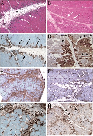
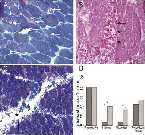
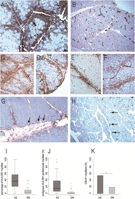
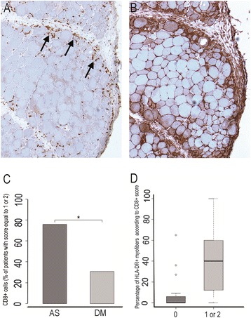
Results: Ubiquitous myofiber HLA-ABC expression was equally observed in ASM and DM (93.9% vs 100%, NS). In contrast, myofiber HLA-DR expression was found in 27/33 (81.8%) ASM (anti-Jo1: 23/26, 88.5%; others: 5/7, 71.4%) vs 4/17 (23.5%) DM patients (p < 0.001). HLA-DR was perifascicular in ASM, a pattern not observed in DM. In addition, C5b-9 deposition was observed on sarcolemma of non-necrotic perifascicular fibers in ASM, while, in DM, C5b-9was mainly detected in endomysial capillaries. CD8 cells were more abundant in ASM than in DM (p < 0.05), and electively located in perimysium or in perifascular endomysium. HLA-DR expression correlated positively with the CD8+ cells infiltrates. Strictly similar observations were made in the confirmatory study.

Conclusion: ASM is characterized by strong myofiber MHC-II/HLA-DR expression with a unique perifascicular pattern, not described so far. HLA-DR detection must be included for routine myopathological diagnosis of inflammatory/dysimmune myopathies. HLA-DR expression in ASM may indicate a specific immune mechanism, possibly involving IFNγ.


Figures

Figure 1
Figure 1
Myopathological features in ASM (A, C, E, G) and DM (B, D, F, H). A: Myofiber injuries in ASM predominate in perifascicular areas (arrows) with the rounded atrophic appearance of regenerating myofibers; inflammatory infiltrates are mainly observed in perimysium and perifascicular endomysium and are associated with fragmentation of perimysium (asterisk). B: Perifascicular atrophy in DM characterized bymyofibers in peripheral regions of fascicles with a cross-sectional area substantially lower than in the rest of the fascicle(arrows). C: Myofiber NCAM expression (arrows) typically restricted to perifascular layers in ASM. D: In DM, NCAM-expressing myofibers are both perifascular (arrows) or grouped in foci (arrowheads). E, F: Diffuse myofiber reexpression of HLA-ABC in ASM (E) and DM (F), with marked perifascicular reinforcement in ASM (E). G, H: Macrophages in perimysium in the vicinity of fascicles (arrows) in ASM (G), and in perimysium (arrows) and perivascular areas (arrowhead) in DM (H). Frozen sections; hematoxylin-eosin (A, B) and immunoperoxidase technique for NCAM (C, D), HLA-ABC (E, F) and CD68 (G, H).
Figure 2
Figure 2
Comparison of myopathological lesions in DM (A, B) and ASM(C). A: Myosinolysis defined by the presence of punched-out vacuoles (arrows) or myofibrillar rarefaction areas, corresponding to foci of myosin filament proteolysis. B: Microinfarcts defined by numerous contiguous fibres within a fascicle, at the same stage of necrosis or regeneration. C: Perimysial fragmentation defined by focal clear areas (asterisk) and spumous appearance of adjacent areas (arrowhead). D: Percentages of cases with presence of the lesion (perimysial fragmentation, microinfarcts, vacuoles, and perifascicular atrophy) in ASM and DM. (*) indicates p < 0.05.Frozen sections (A-C); hematoxylin-eosin (A, B) and Masson trichrome (C).
Figure 3
Figure 3
HLA-DR expression and C5b-9 deposition in ASM and DM. A-F: Regular myofiber HLA-DR expression in perifascicular areas in ASM (A, C-F) contrasting with the lack or very few number of positive myofibers in DM (B); positive staining of endomysial vessels (arrows) is physiological (C). G, H: C5b-9 deposition observed at the surface of perifascicular myofibers in ASM (G) and in endomysial vessel walls (arrows) in DM (H). I,J: Percentage of HLA-DR-positive myofibers (I) and of contiguous HLA-DR-positive perifascicular myofibers (J) in ASM and DM. K: Percentages of cases with presence of C5b-9 deposition at the surface of non-necrotic myofibers in ASM and DM. (*) indicates p < 0.05.Frozen sections (A-H); immunoperoxidase technique for HLA-DR (A-F) and C5b-9 (G, H).
Figure 4
Figure 4
CD8+ T-cell infiltration and myofiber HLA-DR expression. A, B: Comparison of CD8+ T-cells infiltration and myofiber HLA-DR expression in a same case of ASM. CD8+ T-cells (arrows) are detected in perifascicular endomysium and adjacent perimysium (A), in the areas corresponding to those of the strongest myofiber HLA-DR expression (B). Frozen section; immunoperoxidase technique for CD8 and HLA-DR. C: Percentage of patients with a CD8+ score equal to 1 or 2, in ASM and DM; (*) indicates p < 0.05. D: Percentage of HLA-DR-positive myofibers according to the degree of CD8+ T-cell infiltration. Comparison was done between patients with a CD8+ score null and patients with score equal to 1 or 2, ASM and DM patients having been pooled.

References

    1. De Bleecker JL, Lundberg IE, de Visser M, Group EMMBS 193rd ENMC International workshop Pathology diagnosis of idiopathic inflammatory myopathies 30 November - 2 December 2012, Naarden, The Netherlands. Neuromuscul Disord. 2013;23(11):945–951. doi: 10.1016/j.nmd.2013.07.007. - DOI - PubMed
    1. Hoogendijk JE, Amato AA, Lecky BR, Choy EH, Lundberg IE, Rose MR, Vencovsky J, de Visser M, Hughes RA. 119th ENMC international workshop: trial design in adult idiopathic inflammatory myopathies, with the exception of inclusion body myositis, 10–12 October 2003, Naarden, The Netherlands. Neuromuscul Disord. 2004;14(5):337–345. doi: 10.1016/j.nmd.2004.02.006. - DOI - PubMed
    1. Gitiaux C, Kostallari E, Lafuste P, Authier FJ, Christov C, Gherardi RK. Whole microvascular unit deletions in dermatomyositis. Ann Rheum Dis. 2013;72(3):445–452. doi: 10.1136/annrheumdis-2012-201822. - DOI - PubMed
    1. Ranque B, Bérezné A, Le-Guern V, Pagnoux C, Allanore Y, Launay D, Hachulla E, Authier FJ, Gherardi R, Kahan A, Cabane J, Guillevin L, Mouthon L. Myopathies related to systemic sclerosis: a case–control study of associated clinical and immunological features. Scand J Rheumatol. 2010;39(6):498–505. doi: 10.3109/03009741003774626. - DOI - PubMed
    1. Mahler M, Miller FW, Fritzler MJ. Idiopathic inflammatory myopathies and the anti-synthetase syndrome: a comprehensive review. Autoimmun Rev. 2014;13(4–5):367–371. doi: 10.1016/j.autrev.2014.01.022. - DOI - PMC - PubMed

Publication types

Supplementary concepts