

Review
doi: 10.1038/jid.2014.401.
Epub 2014 Oct 23.
Innate lymphoid cells in the skin
Affiliations
- PMID: 25339380
- PMCID: PMC4556524
- DOI: 10.1038/jid.2014.401
Item in Clipboard
Review
Innate lymphoid cells in the skin
J Invest Dermatol.
2015 Mar.
Abstract
Innate lymphoid cells (ILCs) are part of a heterogeneous family of innate immune cells with newly identified roles in mediating immunity, tissue homeostasis, and pathologic inflammation. Here, we review recent studies delineating the roles of ILCs in the pathogenesis of multiple inflammatory skin disorders and their unique effector functions. Finally, we address how these studies have informed our understanding of the regulation of ILCs and the therapeutic potential of targeting these cells in the context of skin inflammation.
Figures
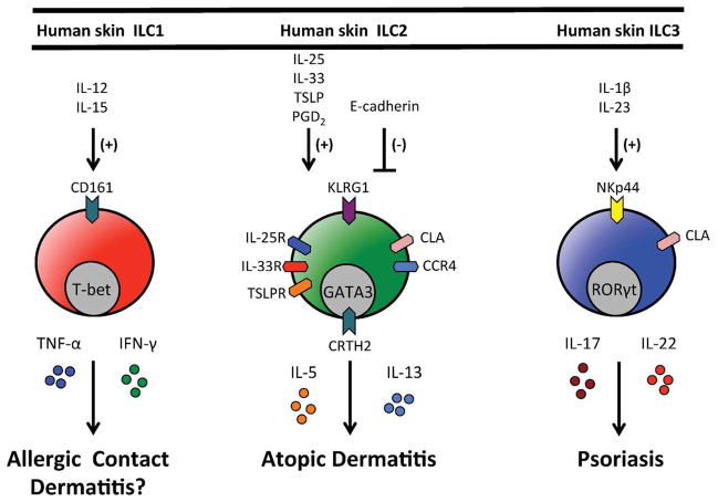
Human skin ILC1s express CD161 and would be predicted to be activated by IL-12 and IL-15 and produce the effector cytokines TNF-α and IFN-γ. Human skin ILC2s express KLRG1, CLA, CCR4, IL-25R (IL-17Rb), IL-33R (ST2), CRTH2 and TSLPR. In response to IL-25, IL-33 and/or TSLP they produce effector cytokines IL-5 and IL-13 and have been implicated in atopic dermatitis. Human skin ILC2s also migrate in response to PGD2. Human skin ILC3s express NKp44 and CLA, produce IL-17 and IL-22 in response to IL-1β and IL-23 and have been implicated in psoriasis.
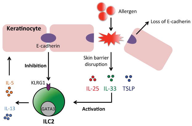
Human skin ILC2s have been implicated in the pathogenesis of atopic dermatitis and are activated by the epithelial cell-derived cytokines IL-25, IL-33 and/or TSLP to produce IL-5 and IL-13. In contrast, the keratinocyte cell adhesion molecule E-cadherin has been shown to inhibit the activation of skin ILC2s, possibly via KLRG1. Atopic dermatitis-associated inflammation and loss of skin barrier protein filaggrin expression is associated with the loss of E-cadherin expression in keratinocytes.
References
Publication types
MeSH terms
Grants and funding
LinkOut - more resources
Full Text Sources
Other Literature Sources
