

The DTI connectivity of the human claustrum
- PMID: 25339630
- PMCID: PMC4324054
- DOI: 10.1002/hbm.22667
The DTI connectivity of the human claustrum
Abstract
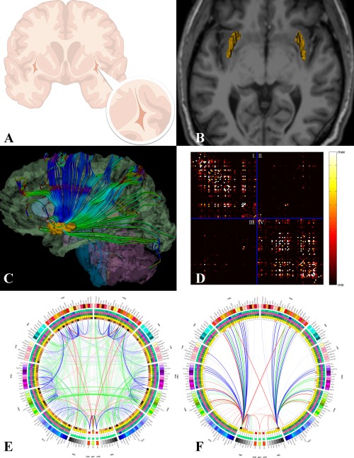
The origin, structure, and function of the claustrum, as well as its role in neural computation, have remained a mystery since its discovery in the 17th century. Assessing the in vivo connectivity of the claustrum may bring forth useful insights with relevance to model the overall functionality of the claustrum itself. Using structural and diffusion tensor neuroimaging in N = 100 healthy subjects, we found that the claustrum has the highest connectivity in the brain by regional volume. Network theoretical analyses revealed that (a) the claustrum is a primary contributor to global brain network architecture, and that (b) significant connectivity dependencies exist between the claustrum, frontal lobe, and cingulate regions. These results illustrate that the claustrum is ideally located within the human central nervous system (CNS) connectome to serve as the putative "gate keeper" of neural information for consciousness awareness. Our findings support and underscore prior theoretical contributions about the involvement of the claustrum in higher cognitive function and its relevance in devastating neurological disease.
Keywords: brain networks; claustrum; cognition; connectivity; consciousness; cortex; diffusion tensor imaging; graph theory; magnetic resonance imaging; neurology.
© 2014 Wiley Periodicals, Inc.
Conflict of interest statement
This research was conducted in absence of any commercial or financial relationships which could be construed as a potential conflict of interest.
Figures
Comment in
-
A giant neuron found wrapped around entire mouse brain.Nature. 2017 Feb 24;543(7643):14-15. doi: 10.1038/nature.2017.21539. Nature. 2017. PMID: 28252090 No abstract available.
References
-
- Arikuni T, Kubota K (1985): Claustral and amygdaloid afferents to the head of the caudate nucleus in macaque monkeys. Neurosci Res 2:239–254. - PubMed
-
- Arnow BA, Desmond JE, Banner LL, Glover GH, Solomon A, Lake Polan M, Lue TF, Atlas SW (2002). Brain activation and sexual arousal in healthy, heterosexual males. Brain 125(Pt 5):1014–1023. - PubMed
-
- Basser PJ, Pajevic S, Pierpaoli C, Duda J, Aldroubi A (2000): In vivo fiber tractography using DT‐MRI data. Magn Reson Med 44:625–632. - PubMed
-
- Bassett DS, Brown JA, Deshpande V, Carlson JM, Grafton ST (2011): Conserved and variable architecture of human white matter connectivity. Neuroimage 54:1262–1279. - PubMed
Publication types
MeSH terms
Grants and funding
LinkOut - more resources
Full Text Sources
Other Literature Sources
