. 2014 Dec 1;193(11):5637-48.
doi: 10.4049/jimmunol.1400606. Epub 2014 Oct 22.

ABCG1 is required for pulmonary B-1 B cell and natural antibody homeostasis

Affiliations

ABCG1 is required for pulmonary B-1 B cell and natural antibody homeostasis

Angel Baldan et al. J Immunol. .

Abstract

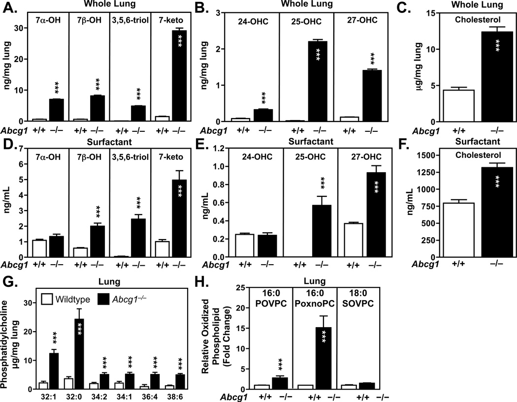
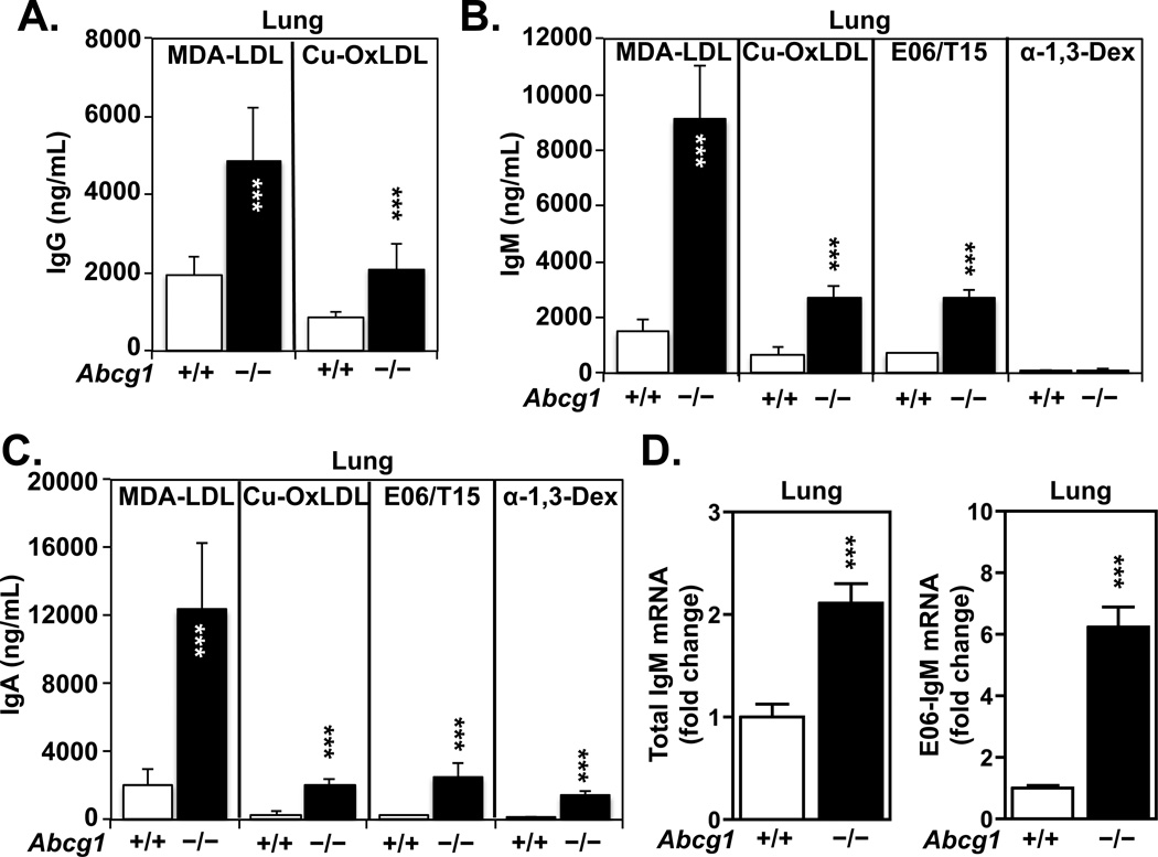
Many metabolic diseases, including atherosclerosis, type 2 diabetes, pulmonary alveolar proteinosis, and obesity, have a chronic inflammatory component involving both innate and adaptive immunity. Mice lacking the ATP-binding cassette transporter G1 (ABCG1) develop chronic inflammation in the lungs, which is associated with the lipid accumulation (cholesterol, cholesterol ester, and phospholipid) and cholesterol crystal deposition that are characteristic of atherosclerotic lesions and pulmonary alveolar proteinosis. In this article, we demonstrate that specific lipids, likely oxidized phospholipids and/or sterols, elicit a lung-specific immune response in Abcg1(-/-) mice. Loss of ABCG1 results in increased levels of specific oxysterols, phosphatidylcholines, and oxidized phospholipids, including 1-palmitoyl-2-(5'-oxovaleroyl)-sn-glycero-3-phosphocholine, in the lungs. Further, we identify a niche-specific increase in natural Ab (NAb)-secreting B-1 B cells in response to this lipid accumulation that is paralleled by increased titers of IgM, IgA, and IgG against oxidation-specific epitopes, such as those on oxidized low-density lipoprotein and malondialdehyde-modified low-density lipoprotein. Finally, we identify a cytokine/chemokine signature that is reflective of increased B cell activation, Ab secretion, and homing. Collectively, these data demonstrate that the accumulation of lipids in Abcg1(-/-) mice induces the specific expansion and localization of B-1 B cells, which secrete NAbs that may help to protect against the development of atherosclerosis. Indeed, despite chronic lipid accumulation and inflammation, hyperlipidemic mice lacking ABCG1 develop smaller atherosclerotic lesions compared with controls. These data also suggest that Abcg1(-/-) mice may represent a new model in which to study the protective functions of B-1 B cells/NAbs and suggest novel targets for pharmacologic intervention and treatment of disease.


Conflict of interest statement

Disclosures

The authors have no conflicts of interest or disclosures.

J.L.W. and X.Q. have patents and disclosures related to the use of oxidation-specific antibodies, which are owned by the University of California San Diego.

Figures

Figure 1
Figure 1. Gating strategy for the isolation of B-1 B cells
(A) Flow cytometry gating strategy to identify B cell subsets in lung, peritoneal cavity, pleural cavity and spleen. Single cell suspensions were stained with different fluorophore-conjugated antibodies and analyzed by flow cytometry. Among single cells, the live cells were selected for further analysis to identify B cells (CD19+), B-1 B cells (CD19+IgM+CD11b+), B-1a B cells (CD19+IgM+CD11b+CD5+), and B-1b B cells (CD19+IgM+CD11b+CD5). (B) Isotype controls for lung B-1 B cells. Single cell suspensions were stained with antigen-specific fluorophore-conjugated antibodies and control fluorephore-conjugated antibodies and analyzed by flow cytometry.
Figure 2
Figure 2. Specific expansion of B-1 B cells in lungs and pleural cavities of Abcg1−/− mice
(A–B) Flow cytometric analysis of cells isolated from the lungs of 6 month old chow-fed wildtype and Abcg1−/− mice. Single cell suspensions were analyzed for expression of CD19 and CD3 as markers for B (A) and T (B) cells, respectively. (C) Representative flow cytometry contour plot of cells obtained from the pleural cavities of 6 month old chow-fed wildtype and Abcg1−/− mice, stained with CD19, CD11b and IgM as markers of B-1 B cells. (D–E) Flow cytometric analysis of cells from lung and pleural cavity (D) or spleen and peritoneal (PerC) cavity (E) from 6 month old chow-fed wildtype and Abcg1−/− mice. Peritoneal and pleural cavities were flushed with saline (5 mL and 3 mL, respectively), and cells collected. Single cell suspensions were analyzed for expression of CD19, CD11b and IgM, as markers for B-1 B cells. (F) Single cell suspensions from the lungs and spleens of 6 month old chow-fed wildtype and Abcg1−/− mice were stained with CD19, CD11b, IgM and CD21 to identify B-2 cells. (G) Single cell suspensions from the pleural cavities and lungs of 6 month old chow-fed wildtype and Abcg1−/− mice were stained with CD19, CD11b and IgM to identify B-1 B cells, and CD5 to distinguish B-1a and B-1b cells. Wildtype (WT); Abcg1−/−(KO) mice. (H–I) Quantification of absolute cell numbers of B-1a and B-1b cells from (G). Data are expressed as mean ±SEM; n = 4 mice/genotype; * p<0.05, ** p<0.01, *** p<0.001.
Figure 3
Figure 3. Lipid-driven B cell expansion in lungs and pleural cavities of Abcg1−/− mice
(A–C) Flow cytometric analysis of cells isolated (as in Figure 1) from the lungs, spleens, peritoneal (PerC) and pleural cavities of either 12 week old chow-fed (A) or 12 week old Western diet-fed (B–C) wildtype and Abcg1−/− mice. Single cell suspensions were analyzed for expression of CD19, CD11b and IgM as markers of B-1 B cells. (D) Representative flow cytometry histogram plot of GFP+ cells obtained from the pleural cavity of wildtype (WT) and Abcg1−/− (KO) mice injected with GFP+ B-1 B cells from wildtype mice expressing the GFP transgene under the control of the chicken β-actin promoter. (E) Flow cytometric analysis of GFP+ B-1 B cells recovered from the lungs, spleens, peritoneal and pleural cavities of 6 month old chow-fed wildtype and Abcg1−/− mice. Data are expressed as mean absolute cell number ±SEM; n = 4–6 mice/genotype; *** p<0.001.
Figure 4
Figure 4. Accumulation of cholesterol, phosphatidylcholine and specific oxidized sterol derivatives and phospholipids in lungs and surfactant of Abcg1−/− mice
Lipids were extracted from lung or surfactant as previously described (31). Cholesterol and oxidized cholesterol derivatives were quantified using MS/MS. (A–C) Auto- (A) and enzymatic (B) oxidation products of cholesterol, and total cholesterol (C) were determined in whole lung of 6 month old chow-fed wildtype and Abcg1−/− mice. (D–F) Auto- (D), and enzymatic (E) oxidation products of cholesterol, and total cholesterol (F) were determined in surfactant isolated by broncho-alveolar lavage (as in (31)) from 6 month old chow-fed wildtype and Abcg1−/− mice. Values are total sterol present per mL surfactant volume recovered. (G–H) Total phosphatidycholine phospholipid (G) and oxidized phospholipid (H) species were determined in whole lung of 6 month old chow-fed wildtype and Abcg1−/− mice by GC-MS and ESI-MS/MS, respectively. Data are expressed as mean ±SEM; n = 5 mice/genotype; *** p<0.001.
Figure 5
Figure 5. ABCG1 regulates B-1 B cell homing
Total RNA was extracted from lungs (25 mg) from 6 month old chow-fed (A, C), 12 week old chow-fed (B) or 12 week old Western diet-fed (B) wildtype and Abcg1−/− mice prior to mRNA quantification of cholesterol 25 hydroxylase (Ch25h), Cyp7b1 and Cxcl13 by quantitative RT-PCR. Values were normalized to 36B4. Data are expressed as mean ±SEM; n = 5 mice/genotype; ** p<0.01. (D) Immunohistochemical analysis of lung from 6 month old chow-fed wildtype and Abcg1−/− mice. Frozen tissue sections (10 µm) were incubated with an antibody to mouse CXCL13. A goat anti-mouse AlexaFluor 488-conjugated secondary antibody used for detection. Sections were counter-stained with DAPI.
Figure 6
Figure 6. Increased immunoglobulins in lungs and plasma of Abcg1−/− mice
(A) Immunohistochemical analysis of lung and spleen from 6 month old chow-fed wildtype and Abcg1−/− mice. Frozen tissue sections (10 µm) were stained with secondary HRP-conjugated antibodies for IgA, IgG, and IgM. Arrows indicate positive staining. (B) Lungs of 6 month old chow-fed wildtype and Abcg1−/− mice were extensively perfused with PBS to remove blood contaminants prior to homogenization and isolation of total lung proteins. Proteins were separated by SDS-PAGE, transferred to nitrocellulose membrane prior to incubation with antibodies to IgA (α chain, 55 kDa), IgG (γ chain, 53 kDa), IgM (µ chain, 65 kDa), and β-actin (42 kDa). (C) Antibodies were extracted from lung tissue (50 mg) following an adapted protocol from Ylä-Herttuala et al. (38). Lung antibodies were diluted 1:10 and then the quantity of total IgA, IgG and IgM determined using ELISA techniques as described in the Methods section. Data (ng/mL) are expressed as mean antibody titer over wildtype levels at a 1:10 dilution. Bars represent mean ±SEM of triplicate determinations of individual mice, comparing total immunoglobulin in Abcg1−/− vs. wildtype mice; n = 4–6 mice/genotype; *** p<0.001. (D) Lung lipid extracts (from 25 mg tissue) from 6 month old chow-fed wildtype and Abcg1−/− mice were spotted onto nitrocellulose membrane, and incubated in the presence of total lung protein extract (from 15 mg tissue) from 6 month old chow-fed wildtype or Abcg1−/− mice. Secondary HRP-conjugated goat anti-mouse antibodies to IgA, IgG, or IgM were used to detect the specific antibody isotype.
Figure 7
Figure 7. Specific lipid antigens and antibodies are present in the lungs ofAbcg1−/− mice
(A–C) Antibodies were extracted from lung tissue as in (Figure 5), diluted 1:10 and tested for binding to the indicated antigens. HRP-conjugated IgG (A), IgM (B), or IgA (C), were used for detection. Data are presented as mean antibody titer (ng/mL) ±SEM comparing Abcg1−/− vs. wildtype mice; n = 4 mice/genotype; *** p<0.001. α-1,3-Dex, α-1,3-Dextran; Cu-Ox, copper oxidized; LDL, low density lipoprotein; MDA, malondialdehyde. (D) Total RNA was extracted from lungs (25 mg tissue) of wildtype and Abcg1−/− mice prior to mRNA quantification of total IgM and E06-IgM by Taqman RT-PCR. Values were normalized to GAPDH. Data are expressed as fold change ±SEM comparing Abcg1−/− vs. wildtype mice; n = 4 mice/genotype; *** p<0.001.
Figure 8
Figure 8. Oxidized lipid antigens are present in lungs and atherosclerotic lesions of Abcg1−/− mice
(A) Frozen lung tissue sections (10 µm) from 6 month old chow-fed wildtype and Abcg1−/− mice were incubated in the presence of mouse E06 antibodies that recognize and bind to oxPL. Secondary anti-mouse Alexafluor 488-conjugated IgM was used for detection. (B) Frozen lung tissue sections from 6 month old chow-fed wildtype and Abcg1−/− mice that were injected with GFP+ cells were incubated in the presence of antibodies that recognize PCNA (proliferative marker) and also analyzed for GFP expression. Secondary anti-rat Alexafluor 594 was used for detection. All tissue sections were counter-stained with DAPI.
Figure 9
Figure 9. Effects of loss of ABCG1 on lipid homeostasis, B-1 B cell homing and natural antibody production
ABCG1 is an intracellular sterol transporter highly expressed in many cell types including B cells, macrophages and pulmonary type II pneumocytes. Loss of ABCG1 from macrophages, particularly in the lung, results in the accumulation of cholesterol and 25-hydroxysterol, and increased levels of the enzymes cholesterol 25-hydroxylase (Ch25h) and Cyp7b1. Increased conversion of 25-hydroxycholesterol to 7α,25-hydroxycholesterol by Cyp7b1 activates the G protein-coupled receptor EBI2/GPR83 on the surface of B cells. Increased secretion of the chemokine CXCL13 from macrophages activates its receptor CXCR5, also expressed on the surface of B cells. This combination of signals results in increased homing of B-1 B cells specifically to the pleural cavity and lungs of Abcg1−/− mice. Loss of ABCG1 from type II pneumocytes results in increased generation of oxidized phospholipids, which cause the stimulation of antibody production by B-1a B cells.

References

    1. Baumgarth N. The double life of a B-1 cell: self-reactivity selects for protective effector functions. Nat. Rev. Immunol. 2011;11:34–46. - PubMed
    1. Hardy RR. B-1 B Cell Development. J. Immunol. 2006;177:2749–2754. - PubMed
    1. Montecino-Rodriguez E, Dorshkind K. Formation of B-1 B Cells from Neonatal B-1 Transitional Cells Exhibits NFkB Redundancy. J. Immunol. 2011;187:5712–5719. - PMC - PubMed
    1. Montecino-Rodriguez E, Dorshkind K. B-1 B cell development in the fetus and adult. Immunity. 2012;36:13–21. - PMC - PubMed
    1. Martin F, Kearney JF. B-cell subsets and the mature ­preimmune repertoire. Marginal zone and B1 B cells as part of a “natural immune memory”. Immunol. Rev. 2000;175:70–79. - PubMed

Publication types

MeSH terms