. 2014 Oct;4(10):140121.
doi: 10.1098/rsob.140121.

Understanding the functional difference between growth arrest-specific protein 6 and protein S: an evolutionary approach

Affiliations

Understanding the functional difference between growth arrest-specific protein 6 and protein S: an evolutionary approach

Romain A Studer et al. Open Biol. 2014 Oct.

Abstract

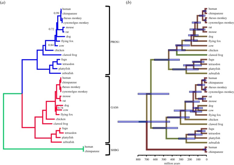
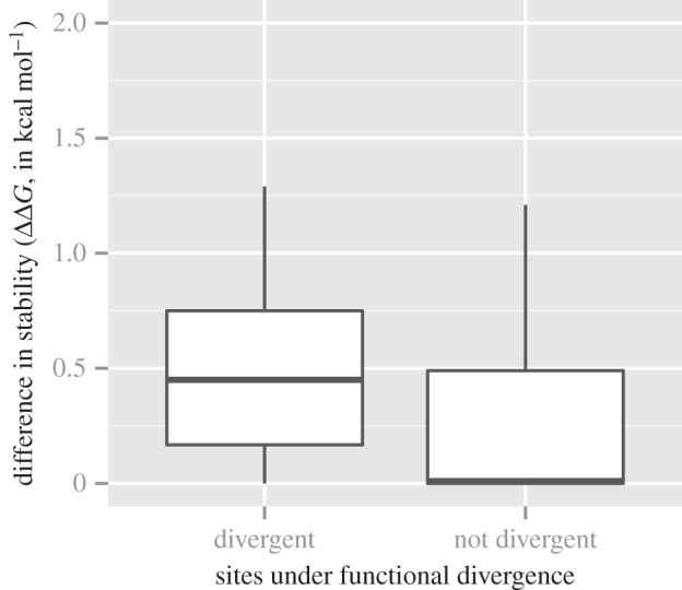
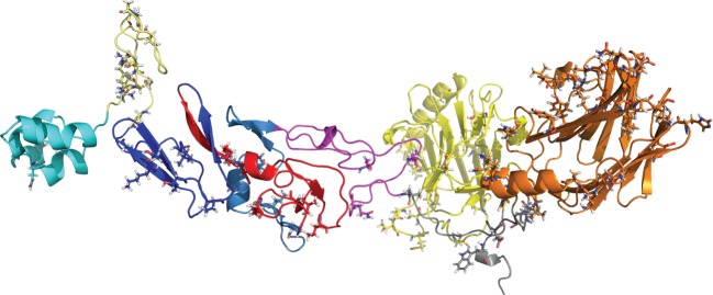
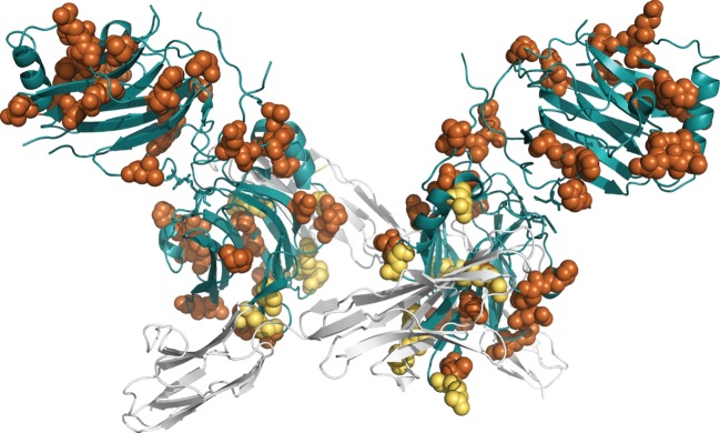
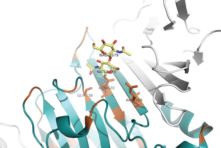
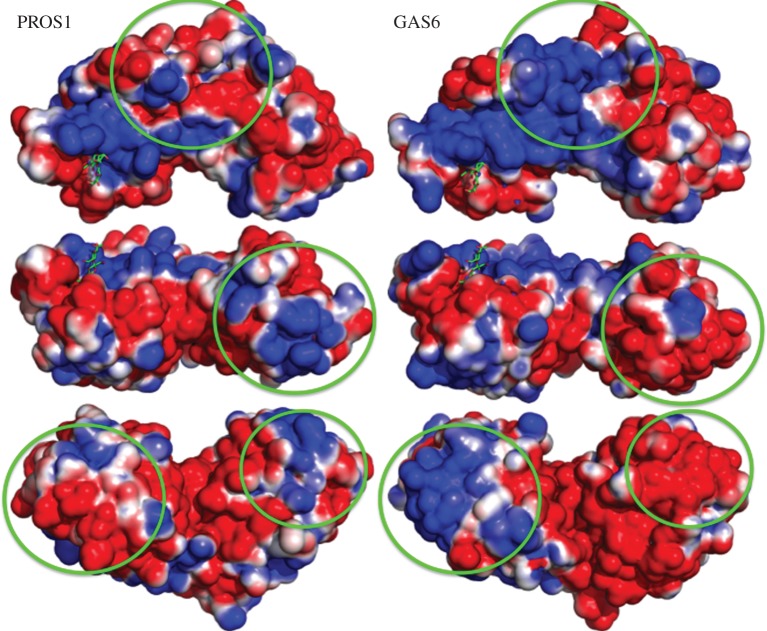
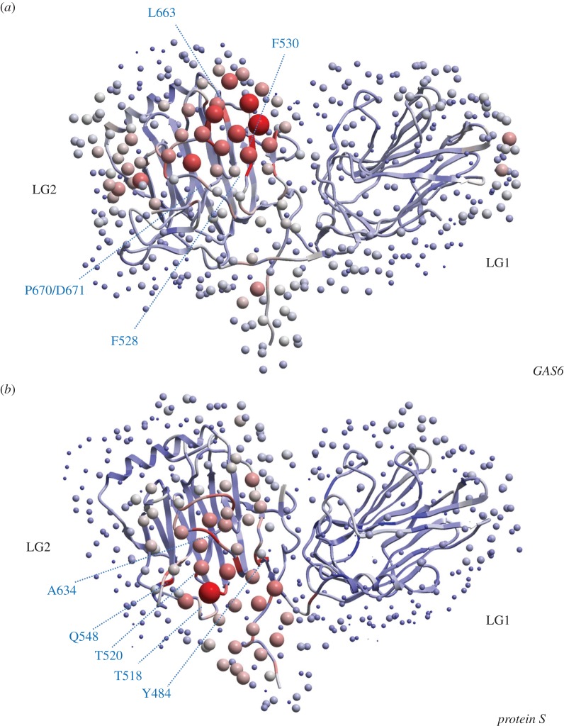
Although protein S (PROS1) and growth arrest-specific protein 6 (GAS6) proteins are homologous with a high degree of structural similarity, they are functionally different. The objectives of this study were to identify the evolutionary origins from which these functional differences arose. Bioinformatics methods were used to estimate the evolutionary divergence time and to detect the amino acid residues under functional divergence between GAS6 and PROS1. The properties of these residues were analysed in the light of their three-dimensional structures, such as their stability effects, the identification of electrostatic patches and the identification potential protein-protein interaction. The divergence between GAS6 and PROS1 probably occurred during the whole-genome duplications in vertebrates. A total of 78 amino acid sites were identified to be under functional divergence. One of these sites, Asn463, is involved in N-glycosylation in GAS6, but is mutated in PROS1, preventing this post-translational modification. Sites experiencing functional divergence tend to express a greater diversity of stabilizing/destabilizing effects than sites that do not experience such functional divergence. Three electrostatic patches in the LG1/LG2 domains were found to differ between GAS6 and PROS1. Finally, a surface responsible for protein-protein interactions was identified. These results may help researchers to analyse disease-causing mutations in the light of evolutionary and structural constraints, and link genetic pathology to clinical phenotypes.

Keywords: evolution; growth arrest-specific protein 6; protein S.


Figures

Figure 1.
Figure 1.
Phylogenetic consensus tree with and without molecular clock of GAS6, PROS1 and SHBG sequences. (a) Tree without a molecular clock model. The GAS6 clade is coloured in red, the PROS1 clade is in blue and the SHBG clade is in green. Values at the nodes indicate posterior probabilities. Only values different from 1.00 are indicated. The lengths of the axes are proportional to the estimated number of mutations per site. (b) Phylogenetic tree under a relaxed clock model. The tree topology is the same as that of the tree in panel (a). The estimated times of divergence of the more important nodes are indicated in electronic supplementary material, table S1. The blue error bars at the nodes represent the 95% confidence limits.
Figure 2.
Figure 2.
Comparison of the stability effect between sites under functional divergence and other sites. The x-axis represents the categories of sites detected by FunDi, BADASP or Selectome. The y-axis represents the absolute median difference in stability effect (ΔΔG, expressed in kcal mol−1) between the group of amino acids in PROS1 versus the group of amino acids in GAS6. These values were estimated with FoldX based on the structure of GAS6 (PDB ID: 2C5D) [22]. Values above 0.5 kcal mol−1 are slightly destabilizing, above 1 kcal mol−1 are destabilizing and above 2 kcal mol−1 are strongly destabilizing.
Figure 3.
Figure 3.
Global view of all sites on PROS1. This is a composite model of the whole PROS1 using different templates. The modelling has been done with Yasara What If. Colouring is domain specific: GLA (cyan), TSR (light yellow), EGF1 (dark blue), EGF2 (red), EGF3 (slate), EGF4 (mangenta), LG1 (yellow) and LG2 (orange).
Figure 4.
Figure 4.
Three-dimensional visualization of GAS6 in complex with Axl (PDB ID: 2C5D). Sites under functional divergence are shown as spheres and coloured in orange. Sites under functional divergence and in contact with Axl (in cartoon and in white) are in yellow. α-helices and β-sheets of GAS6 domain are in blue.
Figure 5.
Figure 5.
Visualization of N-acetylglucosamine (NAG) binding site. The asparagine at position Asn463(420) in GAS6 is mutated to a lysine in PROS1. NAG ligand is in stick and coloured in yellow. Sites under functional divergence are coloured in orange. α-helices and β-sheets of GAS6 domain are in blue. Axl domains are in grey.
Figure 6.
Figure 6.
Visualization of electrostatic surfaces on the SHBG-domain of PROS1 and GAS6. To make the direct comparison between GAS6 and PROS1 easier, we have modelled their SHBG domains using the GAS6 PDB structure (PDB ID: 2C5D). NAG ligand has been added to identify its putative binding pocket. While it is crystallized in GAS6, there is no evidence to indicate whether it can be present in PROS1. Basic surfaces are in blue while acidic surfaces are in red. The NAG ligand is in green. The green circles indicate the observed differences in electrostatic surface potential between GAS6 and PROS1.
Figure 7.
Figure 7.
Optimal docking area analysis for LG1 and LG2-domains of GAS6 and PROS1. Red dots indicate likely interaction areas, blue indicates protein–protein interactions to be unlikely. (a) For GAS6, the most likely interactions are with F528, F530, L663, P670 and D671. (b) For PROS1, the most likely interactions are with Y484, T518, T520, Q548 and A634.

References

    1. Manfioletti G, Brancolini C, Avanzi G, Schneider C. 1993. The protein encoded by a growth arrest-specific gene (gas6) is a new member of the vitamin K-dependent proteins related to protein S, a negative coregulator in the blood coagulation cascade. Mol. Cell Biol. 13, 4976–4985. - PMC - PubMed
    1. Hafizi S, Dahlbäck B. 2006. Gas6 and protein S. Vitamin K-dependent ligands for the Axl receptor tyrosine kinase subfamily. FEBS J. 273, 5231–5244. (doi:10.1111/j.1742-4658.2006.05529.x) - DOI - PubMed
    1. Walker FJ, Chavin SI, Fay PJ. 1987. Inactivation of factor VIII by activated protein C and protein S. Arch. Biochem. Biophys. 252, 322–328. (doi:10.1016/0003-9861(87)90037-3) - DOI - PubMed
    1. Walker FJ. 1980. Regulation of activated protein C by a new protein. A possible function for bovine protein S. J. Biol. Chem. 255, 5521–5524. - PubMed
    1. Hackeng TM, Seré KM, Tans G, Rosing J. 2006. Protein S stimulates inhibition of the tissue factor pathway by tissue factor pathway inhibitor. Proc. Natl Acad. Sci. USA 103, 3106–3111. (doi:10.1073/pnas.0504240103) - DOI - PMC - PubMed

MeSH terms