. 2014 Oct 22;34(43):14210-8.
doi: 10.1523/JNEUROSCI.0326-14.2014.

Regulation of presynaptic Ca2+, synaptic plasticity and contextual fear conditioning by a N-terminal β-amyloid fragment

Affiliations

Regulation of presynaptic Ca2+, synaptic plasticity and contextual fear conditioning by a N-terminal β-amyloid fragment

James L M Lawrence et al. J Neurosci. .

Abstract

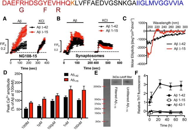
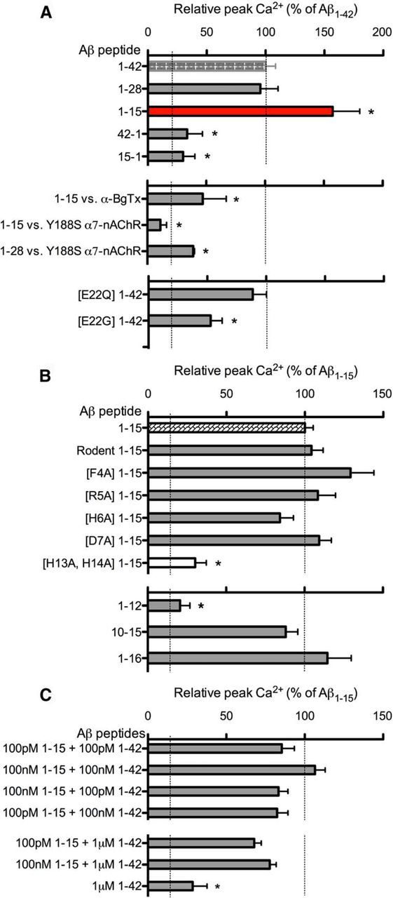
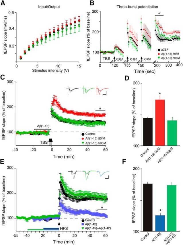
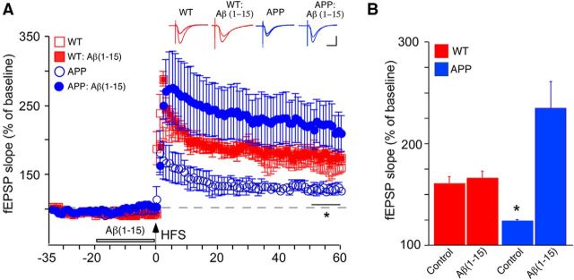
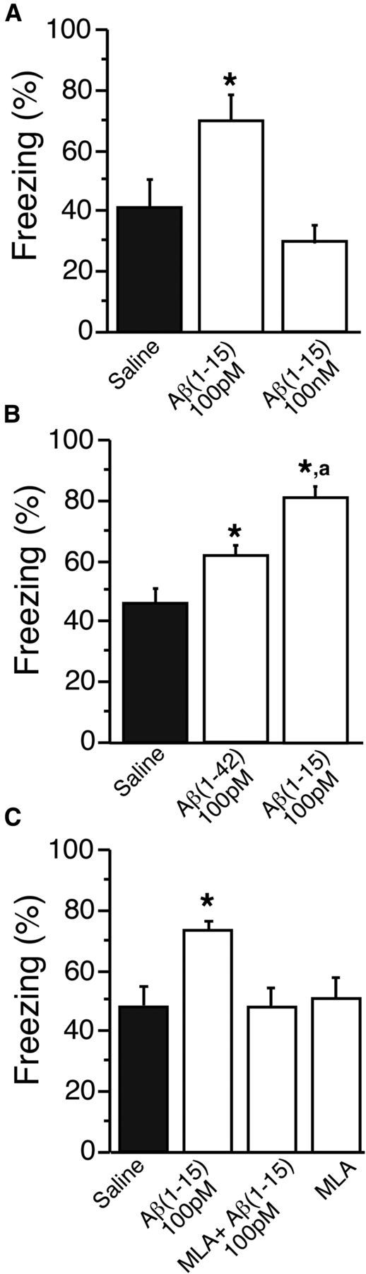
Soluble β-amyloid has been shown to regulate presynaptic Ca(2+) and synaptic plasticity. In particular, picomolar β-amyloid was found to have an agonist-like action on presynaptic nicotinic receptors and to augment long-term potentiation (LTP) in a manner dependent upon nicotinic receptors. Here, we report that a functional N-terminal domain exists within β-amyloid for its agonist-like activity. This sequence corresponds to a N-terminal fragment generated by the combined action of α- and β-secretases, and resident carboxypeptidase. The N-terminal β-amyloid fragment is present in the brains and CSF of healthy adults as well as in Alzheimer's patients. Unlike full-length β-amyloid, the N-terminal β-amyloid fragment is monomeric and nontoxic. In Ca(2+) imaging studies using a model reconstituted rodent neuroblastoma cell line and isolated mouse nerve terminals, the N-terminal β-amyloid fragment proved to be highly potent and more effective than full-length β-amyloid in its agonist-like action on nicotinic receptors. In addition, the N-terminal β-amyloid fragment augmented theta burst-induced post-tetanic potentiation and LTP in mouse hippocampal slices. The N-terminal fragment also rescued LTP inhibited by elevated levels of full-length β-amyloid. Contextual fear conditioning was also strongly augmented following bilateral injection of N-terminal β-amyloid fragment into the dorsal hippocampi of intact mice. The fragment-induced augmentation of fear conditioning was attenuated by coadministration of nicotinic antagonist. The activity of the N-terminal β-amyloid fragment appears to reside largely in a sequence surrounding a putative metal binding site, YEVHHQ. These findings suggest that the N-terminal β-amyloid fragment may serve as a potent and effective endogenous neuromodulator.

Keywords: Ca regulation; fear memory; neuromodulation; synaptic plasticity; β-amyloid.


Figures

Figure 1.
Figure 1.
A N-terminal Aβ fragment encompassing residues 1–15 is a potent and highly effective agonist at α7-nAChRs on presynaptic-like axonal varicosities. A, B, Averaged [Ca2+]i responses (F/F0) in varicosities of NG108–15 cells expressing α7-nAChRs (A) or mouse hippocampal synaptosomes (B) to 100 nm1–42 (A, n = 49; B, n = 16) were compared with responses to 100 nm1–15 (A: n = 178; p < 0.05; B: n = 26; NS), followed sequentially by K+-induced depolarization. Time-series traces are means ± SEM at individual time points. The Ca2+ responses to 100 nm1–42 are similar in magnitude to that observed with nicotine (data not shown; see Khan et al., 2010). C, Representative CD spectra for Aβ1–42 and Aβ1–15. D, Averaged peak Ca2+ responses in varicosities expressing α7-nAChRs to 100 fm (n = 22 or n = 33), 1 pm (n = 26 or n = 21), 100 pm (n = 19 or n = 32), 1 nm (n = 30 or n = 15), and 100 nm (n = 44 or n = 29) Aβ1–42 or Aβ1–15, respectively. All pairs (Aβ1–42 vs Aβ1–15) are significantly different (p < 0.05, t test). E, 4–20% gradient Tris-Tricine PAGE of Aβ1–15 (2 nmol). Positions of molecular weight standards (data not shown) are as marked in kilodaltons. Insets show a comparison of Aβ1–15 (molecular weight, 1827 kDa) before and after (from whole lane) filtration through an Amicon 3 kDa cutoff filter. Dimers and larger oligomers will be excluded by the 3 kDa filter. F, Fibril–aggregate formation of Aβ1–42, Aβ1–15, or Aβ42–1 control peptide, each at 200 nm, assessed in triplicate by a fluorimetric thioflavin (ThT) assay.
Figure 2.
Figure 2.
Structure–function analysis of the N-terminal Aβ domain and fragment. A, Averaged peak Ca2+ responses in varicosities of NG108–15 cells expressing α7-nAChRs to 100 nm1–42 (n = 44), Aβ1–28 (n = 10), and Aβ1–15 (n = 29), and the control peptides Aβ42–1 (n = 24) and Aβ15–1 (n = 38). Peak Ca2+ responses to Aβ1–15 in the presence of 50 nm α-bungarotoxin (α-BgTx, n = 12), Aβ1–15 (n = 20), and Aβ1–28 (n = 11) on Y188S α7-nAChRs. Peak Ca2+ responses to E22Q Aβ1–42 (n = 29) and E22G Aβ1–42 (n = 19). B, Peak Ca2+ responses via α7-nAChRs to rodent Aβ1–42 (n = 49), rodent Aβ1–15 (n = 39), F4A Aβ1–15 (n = 27), R5A Aβ1–15 (n = 46), H6A Aβ1–15 (n = 34), D7A Aβ1–15 (n = 40); H13A/H14A Aβ1–15 (n = 25), Aβ1–12 (n = 48), Aβ1–16 (n = 51), and Aβ10–15 (n = 70). C, Averaged peak Ca2+ responses to mixtures of Aβ1–15 and Aβ1–42 at various concentrations, as noted. *p < 0.05 (Bonferroni post hoc tests) NB. Dashed lines indicate the baseline (background) and respective average maximal responses for either Aβ1–42 and Aβ1–15, as indicated.
Figure 3.
Figure 3.
N-terminal Aβ fragment augments LTP in hippocampal slices in the absence or presence of elevated full-length Aβ. Hippocampal slices were superfused with aCSF containing vehicle (control) or various concentrations of Aβ1–15 without (B–D) or with (E, F) Aβ1–42, followed by the induction of LTP in the CA1 region via theta burst stimulation (TBP: four trains of 100 Hz pulses delivered at 5 Hz repeated three times every 15 s for a total of 3 bursts) or HFS (two 1 s trains of 100 Hz separated by 20 s) through the Schaffer collaterals and expressed as normalized fEPSP slope values. A, Control input/output curves, before treatment. B, Recording during and after the theta burst following 57 fm, 57 pm, or 100 nm1–15 for 20 min, with the start of each burst marked with an arrow. Note the change in time scale (dashed lines) for the bursts: PTP marked with a solid bar. C, TBP-induced LTP with color-coded insets showing example fEPSPs for control aCSF (black), femtomolar Aβ1–15 (red) or picomolar Aβ1–15 (green) for baseline and LTP. The period of Aβ1–15 pretreatment is marked by the open bar. D, Average fEPSP slope values for the end of the plateau (50–60 min post-tetanus), as noted by the solid bar in C (*). E, HFS-induced LTP, with color-coded insets showing example fEPSPs for control aCSF (black), 500 nm1–42 (blue), or 500 nm1–15 followed by 500 nm1–42 (green) for baseline and LTP; periods of peptide pretreatment are marked by the bars. F, Average fEPSP slope values for the end of the plateau (50–60 min post-tetanus), as noted by the solid black bar in E (*). Data are the means ± SD, n = 6 slices/group derived from three experiments. Calibration: horizontal, 10 ms; vertical, 0.4 mV. *p < 0.05 (Bonferroni post hoc tests).
Figure 4.
Figure 4.
N-terminal Aβ fragment rescues LTP deficits in APPswe mouse hippocampal slices. Hippocampal slices from APPswe or wild-type (WT) littermates were superfused with aCSF containing vehicle (Control) or 500 nm1–15. A, HFS-induced LTP, with color-coded insets showing example fEPSPs for slices from WT mice without or with pretreatment with Aβ1–15 (red) or slices from APPswe mice without or with pretreatment with 500 nm1–42 (blue) for baseline and LTP; period of Aβ1–15 pretreatment marked by the open bar. B, Average fEPSP slope values for the end of the plateau (50–60 min post-tetanus), as noted by the solid bar in A (*). Data are the means ± SD, n = 4 slices/group. Calibration: horizontal, 10 ms; vertical, 0.4 mV. *p < 0.05 (Bonferroni post hoc tests).
Figure 5.
Figure 5.
Bilateral delivery of picomolar N-terminal Aβ fragment into the dorsal hippocampus enhances contextual fear conditioning. Mice were trained for contextual fear conditioning under a single-trial paradigm using mild shock. A–C, Twenty-four hours before testing, 1.83 ng/L (100 pm) Aβ1–15 or 1.83 μg/L (100 nm) Aβ1–15 (A); 4.5 ng/L (100 pm) Aβ1–42 or 1.83 ng/L (100 pm) Aβ1–15 (B); and 1.83 ng/L (100 pm) Aβ1–15, 1.83 ng/L (100 pm) Aβ1–15 + 100 nm MLA or 100 nm MLA (C); or sterile saline were bilaterally injected into the dorsal hippocampi. Freezing was measured via TSE videotracking software. Conditioned freezing was assessed by two trained observers. Data are the means ± SEM, n = 6–9 mice/group. *p < 0.005 compared with saline control; ap < 0.05 comparing Aβ1–15 to Aβ1–42 (Bonferroni post hoc tests).

References

    1. Abramov E, Dolev I, Fogel H, Ciccotosto GD, Ruff E, Slutsky I. Amyloid-β as a positive endogenous regulator of release probability at hippocampal synapses. Nat Neurosci. 2009;12:1567–1576. doi: 10.1038/nn.2433. - DOI - PubMed
    1. Bell KA, O'Riordan KJ, Sweatt JD, Dineley KT. MAPK recruitment by β-amyloid in organotypic hippocampal slice cultures depends on physical state and exposure time. J Neurochem. 2004;91:349–361. doi: 10.1111/j.1471-4159.2004.02722.x. - DOI - PubMed
    1. Bellinger FP, Wilce PA, Bedi KS, Wilson P. Long-lasting synaptic modification in the rat hippocampus resulting from NMDA receptor blockade during development. Synapse. 2002;43:95–101. doi: 10.1002/syn.10020. - DOI - PubMed
    1. Buée L, Ding W, Delacourte A, Fillit H. Binding of secreted human neuroblastoma proteoglycans to the Alzheimer's amyloid A4 peptide. Brain Res. 1993;601:154–163. doi: 10.1016/0006-8993(93)91706-X. - DOI - PubMed
    1. Chin JH, Ma L, MacTavish D, Jhamandas JH. Amyloid β protein modulates glutamate-mediated neurotransmission in the rat basal forebrain: involvement of presynaptic neuronal nicotinic acetylcholine and metabotropic glutamate receptors. J Neurosci. 2007;27:9262–9269. doi: 10.1523/JNEUROSCI.1843-07.2007. - DOI - PMC - PubMed

Publication types

MeSH terms