. 2014 Oct 22;34(43):14243-51.
doi: 10.1523/JNEUROSCI.2283-14.2014.

Effects of HIV-1 Tat on enteric neuropathogenesis

Affiliations

Effects of HIV-1 Tat on enteric neuropathogenesis

Joy Ngwainmbi et al. J Neurosci. .

Abstract

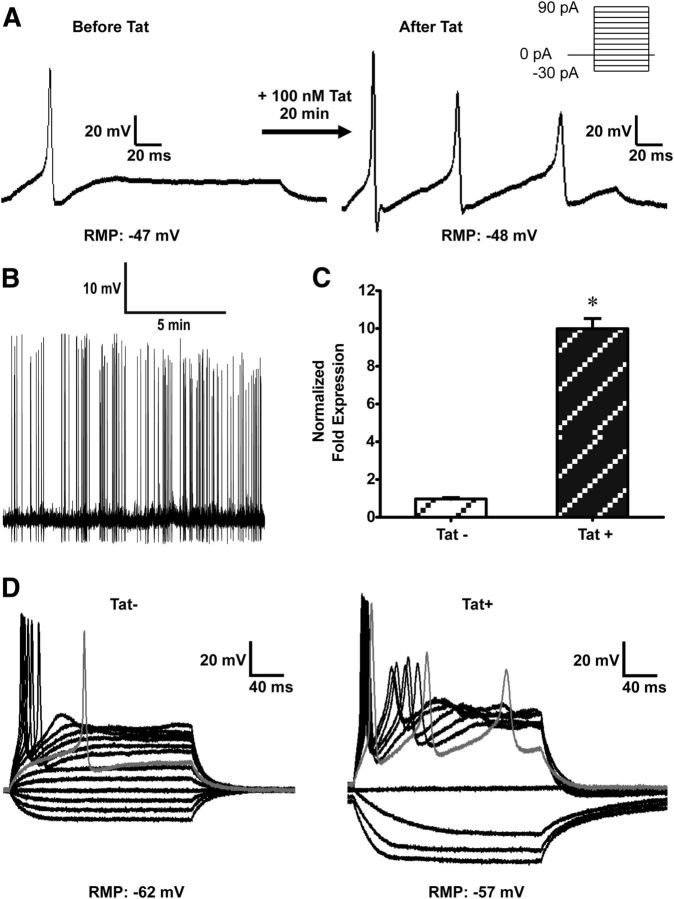
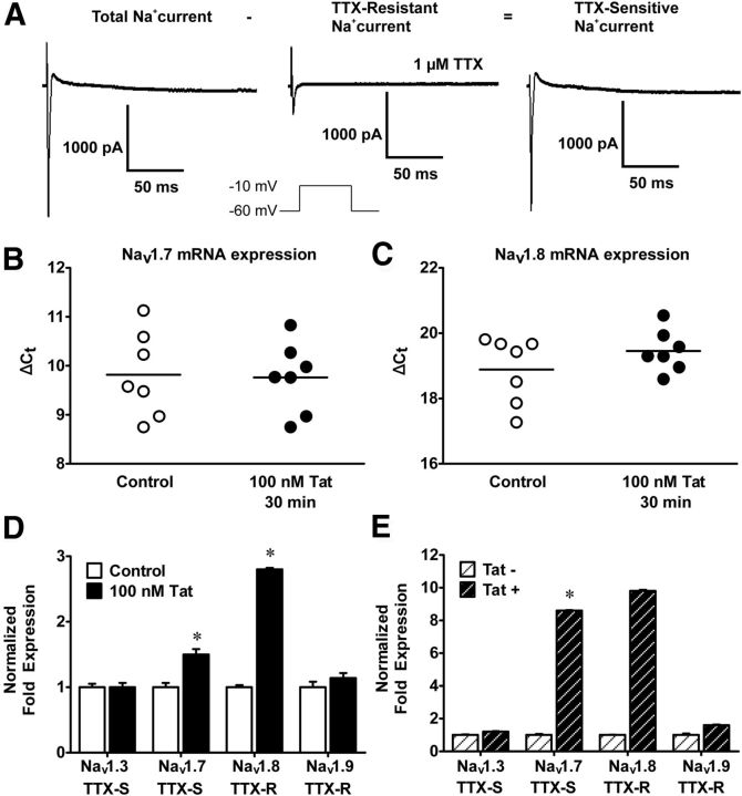
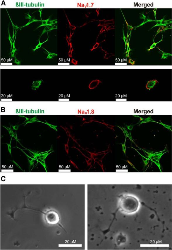
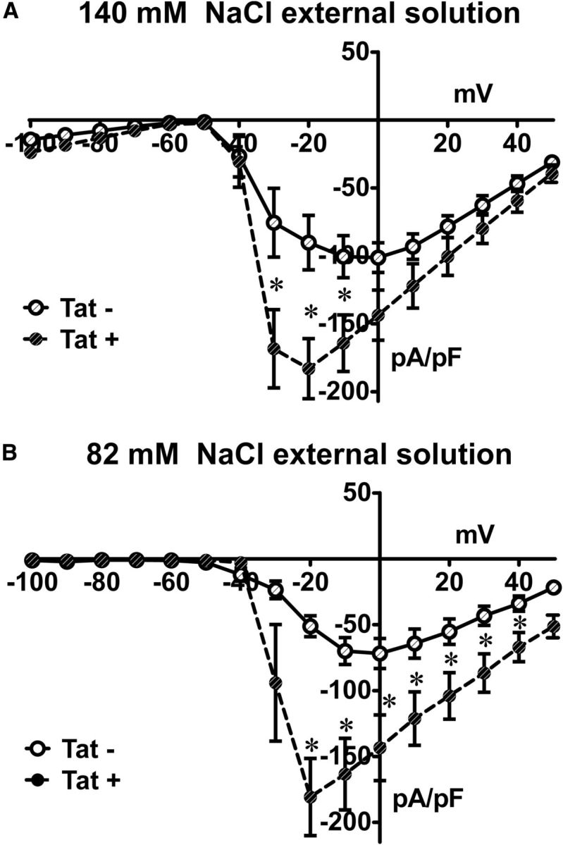
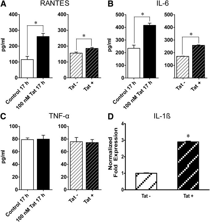
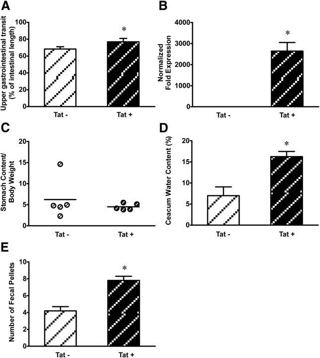
The gastrointestinal (GI) tract presents a major site of immune modulation by HIV, resulting in significant morbidity. Most GI processes affected during HIV infection are regulated by the enteric nervous system. HIV has been identified in GI histologic specimens in up to 40% of patients, and the presence of viral proteins, including the trans-activator of transcription (Tat), has been reported in the gut indicating that HIV itself may be an indirect gut pathogen. Little is known of how Tat affects the enteric nervous system. Here we investigated the effects of the Tat protein on enteric neuronal excitability, proinflammatory cytokine release, and its overall effect on GI motility. Direct application of Tat (100 nm) increased the number of action potentials and reduced the threshold for action potential initiation in isolated myenteric neurons. This effect persisted in neurons pretreated with Tat for 3 d (19 of 20) and in neurons isolated from Tat(+) (Tat-expressing) transgenic mice. Tat increased sodium channel isoforms Nav1.7 and Nav1.8 levels. This increase was accompanied by an increase in sodium current density and a leftward shift in the sodium channel activation voltage. RANTES, IL-6, and IL-1β, but not TNF-α, were enhanced by Tat. Intestinal transit and cecal water content were also significantly higher in Tat(+) transgenic mice than Tat(-) littermates (controls). Together, these findings show that Tat has a direct and persistent effect on enteric neuronal excitability, and together with its effect on proinflammatory cytokines, regulates gut motility, thereby contributing to GI dysmotilities reported in HIV patients.

Keywords: AIDS; HIV; cytokines; myenteric; sodium channels.


Figures

Figure 1.
Figure 1.
Tat increased enteric neuronal excitability. A, Representative traces showing current-clamp recordings of a neuron in the absence and in the presence of 100 nm Tat. Increased neuronal excitability in response to 100 nm Tat is evidenced by an increase in the number of action potentials evoked. Action potentials were initiated after 30 pA current injection in the absence of Tat and 10 pA after continuous perfusion with 100 nm Tat; n = 5. B, Spontaneous action potentials were recorded in 4 neurons after Tat exposure. C, RT-PCR experiments showing that tat gene is expressed in Tat+ LMMP and absent in Tat LMMP. *p < 0.05 (t test). D, Neurons isolated from Tat+ transgenic mice were also more excitable than Tat mice. Gray line indicates response recorded at rheobase.
Figure 2.
Figure 2.
Tat changes the steady-state voltage dependence of activation of sodium channels. Na+ channel activation/inactivation was examined using whole-cell patch-clamp experiments in voltage-clamp mode and a double-pulse protocol in the presence and absence of 100 nm Tat and without Tat (control). Cs+ is present in the internal solution to block outward K+ current. A, Current density/voltage curve of controls (untreated cells) and Tat-pretreated neurons. B, Boltzmann curve analysis of inactivation and activation of Na+ indicates a leftward shift of the activation curve in response to Tat. C, Significant difference in V0.5 of activation. D, There was no significant difference in V0.5 of inactivation. *p < 0.05 (t test).
Figure 3.
Figure 3.
Tat transcriptionally modulates Nav1.7 and Nav1.8. A, Representative raw traces of voltage-clamp experiments showing the presence of TTX-sensitive and -resistant sodium channels in neurons isolated from the adult mouse ileum. B, C, Quantitative PCR of LMMP preparations after 30 min of Tat treatment. Nav1.7 and Nav1.8 mRNA levels are unchanged. D, After long-term exposure (2–3 d) to Tat, Nav1.7 and Nav1.8 mRNA levels are significantly increased, but Nav1.3 and Nav1.9 levels are not affected by Tat pretreatment. E, Significantly higher Nav1.7 and Nav1.8 mRNA are observed in Tat+ compared with Tat LMMPs. *p < 0.05 (ANOVA followed by Bonferroni's post hoc test).
Figure 4.
Figure 4.
Nav1.7 and Nav1.8 are expressed on enteric neurons and are upregulated by Tat. Representative immunocytochemistry showing β-III tubulin (green), a neuronal marker, and the sodium channel isoforms (red) Nav1.7 (A) and Nav1.8 (B). Sodium channel isoforms colocalize with neurons and are associated with the cell membrane. C, Phase-contrast microscopic images showing neuronal cells with rounded cell bodies used for patch-clamp analysis.
Figure 5.
Figure 5.
Tat+ neurons had higher sodium current densities compared with Tat. Current density/voltage curves of Tat+ and Tat neurons at 140 mm (A) and 82 mm (B) bath solution NaCl concentration. Tat+ neurons had higher sodium current densities. *p < 0.05 (t test).
Figure 6.
Figure 6.
Tat selectively upregulates proinflammatory cytokines. ELISA assay, assessing proinflammatory cytokine release. A–C, Tat increases RANTES and IL-6 levels but has no effect on TNF-α levels. D, PCR showing increased levels of IL-1β mRNA. *p < 0.05 (Student's t test).
Figure 7.
Figure 7.
A, Tat increased GI motility in Tat transgenic mice. Tat+ mice showed higher upper GI transit rates compared with Tat. GI transit was measured as distance traveled by charcoal meal. Quantitative PCR showing expression of Tat following 1 week of DOX treatment followed by 3 weeks of regular chow diet. B, Tat was expressed in Tat+ and absent in Tat mouse ilea after 3 weeks without DOX. C, Gastric emptying shows no significant difference in stomach stasis rates between Tat+ and Tat mice. D, Cecal water content is significantly higher in Tat+ mice than in Tat. E, The number of stool pellets was significantly higher in Tat+ than Tat mice. *p < 0.05 (Student's t test).

Similar articles

Cited by

References

    1. Akbarali HI, Giles WR. Ca2+ and Ca(2+)-activated Cl− currents in rabbit oesophageal smooth muscle. J Physiol. 1993;460:117–133. - PMC - PubMed
    1. Anitha M, Vijay-Kumar M, Sitaraman SV, Gewirtz AT, Srinivasan S. Gut microbial products regulate murine gastrointestinal motility via toll-like receptor 4 signaling. Gastroenterology. 2012;143:1006–1016. doi: 10.1053/j.gastro.2012.06.034. - DOI - PMC - PubMed
    1. Asakawa A, Inui A, Kaga T, Katsuura G, Fujimiya M, Fujino MA, Kasuga M. Antagonism of ghrelin receptor reduces food intake and body weight gain in mice. Gut. 2003;52:947–952. doi: 10.1136/gut.52.7.947. - DOI - PMC - PubMed
    1. Brailoiu GC, Brailoiu E, Chang JK, Dun NJ. Excitatory effects of human immunodeficiency virus 1 tat on cultured rat cerebral cortical neurons. Neuroscience. 2008;151:701–710. doi: 10.1016/j.neuroscience.2007.11.031. - DOI - PMC - PubMed
    1. Brenchley JM. Mucosal immunity in human and simian immunodeficiency lentivirus infections. Mucosal Immunol. 2013;6:657–665. doi: 10.1038/mi.2013.15. - DOI - PMC - PubMed

Publication types

MeSH terms

Substances

LinkOut - more resources