. 2014 Oct 22;34(43):14375-87.
doi: 10.1523/JNEUROSCI.1222-14.2014.

PGC-1α provides a transcriptional framework for synchronous neurotransmitter release from parvalbumin-positive interneurons

Affiliations

PGC-1α provides a transcriptional framework for synchronous neurotransmitter release from parvalbumin-positive interneurons

Elizabeth K Lucas et al. J Neurosci. .

Abstract

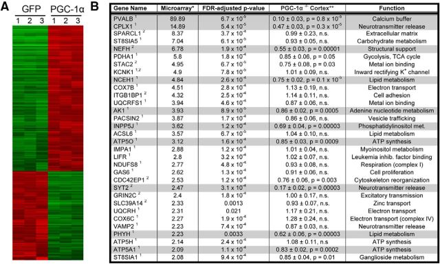
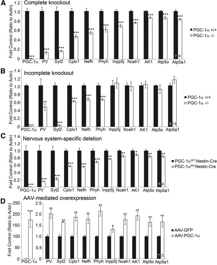
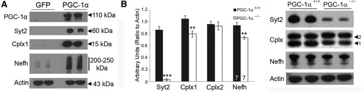
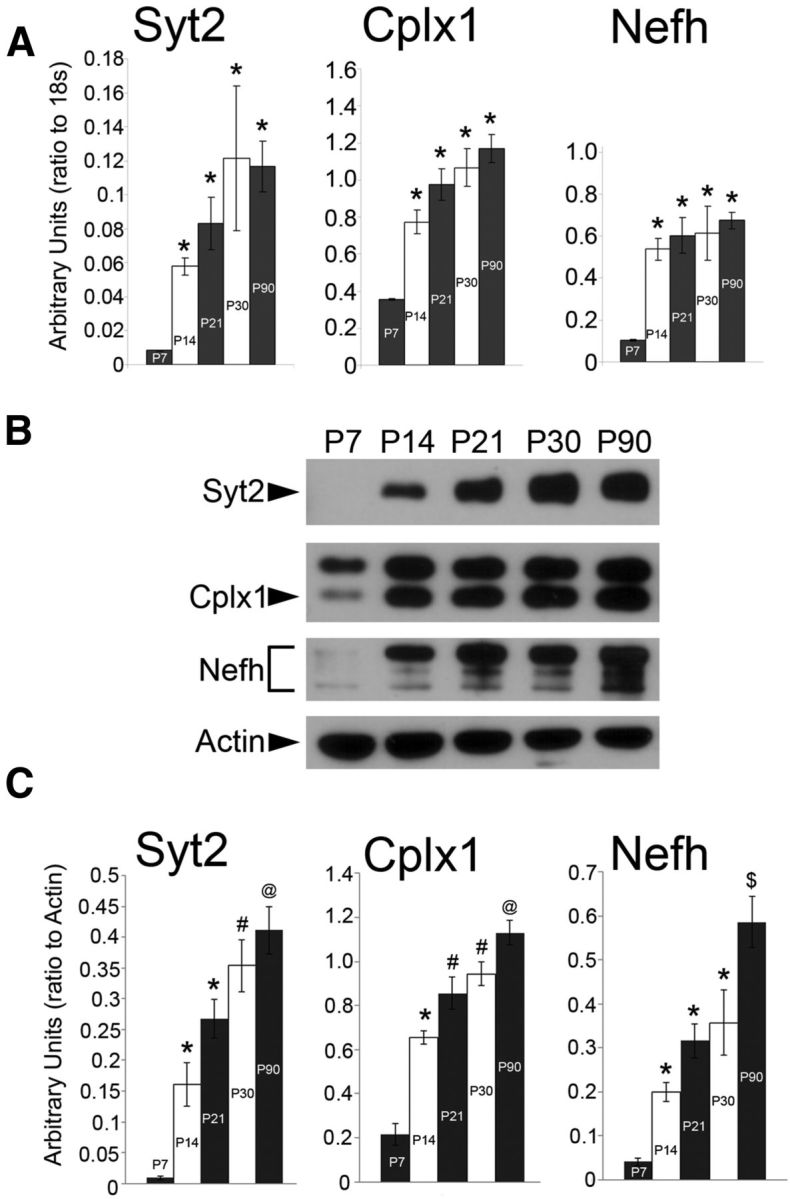
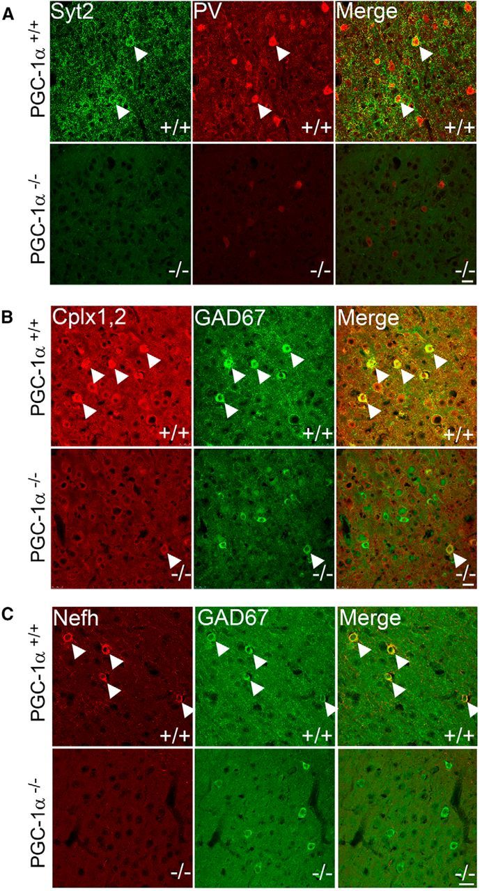
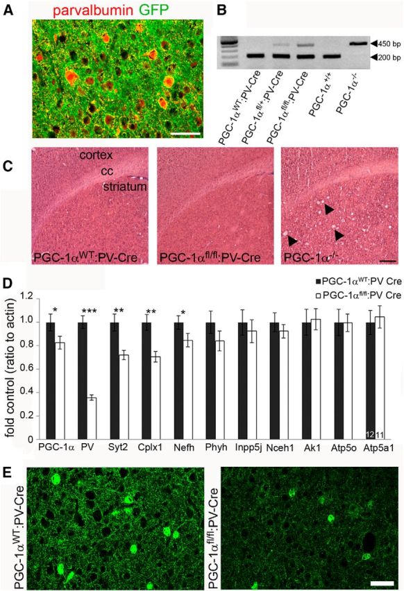
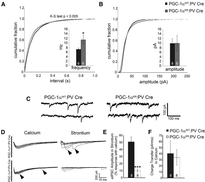
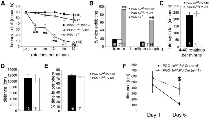
Accumulating evidence strongly implicates the transcriptional coactivator peroxisome proliferator-activated receptor γ coactivator 1α (PGC-1α) in the pathophysiology of multiple neurological disorders, but the downstream gene targets of PGC-1α in the brain have remained enigmatic. Previous data demonstrate that PGC-1α is primarily concentrated in inhibitory neurons and that PGC-1α is required for the expression of the interneuron-specific Ca(2+)-binding protein parvalbumin (PV) throughout the cortex. To identify other possible transcriptional targets of PGC-1α in neural tissue, we conducted a microarray on neuroblastoma cells overexpressing PGC-1α, mined results for genes with physiological relevance to interneurons, and measured cortical gene and protein expression of these genes in mice with underexpression and overexpression of PGC-1α. We observed bidirectional regulation of novel PGC-1α-dependent transcripts spanning synaptic [synaptotagmin 2 (Syt2) and complexin 1 (Cplx1)], structural [neurofilament heavy chain (Nefh)], and metabolic [neutral cholesterol ester hydrolase 1 (Nceh1), adenylate kinase 1 (Ak1), inositol polyphosphate 5-phosphatase J (Inpp5j), ATP synthase mitochondrial F1 complex O subunit (Atp5o), phytanol-CoA-2hydroxylase (Phyh), and ATP synthase mitrochondrial F1 complex α subunit 1 (Atp5a1)] functions. The neuron-specific genes Syt2, Cplx1, and Nefh were developmentally upregulated in an expression pattern consistent with that of PGC-1α and were expressed in cortical interneurons. Conditional deletion of PGC-1α in PV-positive neurons significantly decreased cortical transcript expression of these genes, promoted asynchronous GABA release, and impaired long-term memory. Collectively, these data demonstrate that PGC-1α is required for normal PV-positive interneuron function and that loss of PGC-1α in this interneuron subpopulation could contribute to cortical dysfunction in disease states.

Keywords: Barnes maze; Huntington disease; cortical development; ppargc1a; schizophrenia; strontium.


Figures

Figure 1.
Figure 1.
Identification of novel PGC-1α-dependent genes in neural tissue. A, Microarray heat map of neuroblastoma cells overexpressing PGC-1α and GFP in tandem or control samples overexpressing GFP alone. Red/green indicates an increase/decrease in gene expression of the ∼27,000 tested transcripts. n = 3 per group. B, Microarray data were mined for significantly increased genes with physiological relevance to interneurons based on their neuroanatomical overlap with (1) PGC-1α or (2) PV (indicated by superscript number behind gene name; see Materials and Methods). Selected transcripts were then measured by qRT-PCR in cortical homogenates from PGC-1α+/+ and PGC-1α−/− mice with the hypothesis that a true PGC-1α-dependent gene would be both increased by overexpression and decreased by deletion. Two-tailed t tests with Bonferroni's correction for multiple testing. The gray highlight indicates statistically significant decreases. n = 6–7 per group. *Values expressed as PGC-1α:GFP ratio; **values expressed as PGC-1α−/−/PGC-1α+/+ ratio after normalization to β-actin. Data are presented as mean ± SEM.
Figure 2.
Figure 2.
Regulation of novel transcripts by ablation and overexpression of PGC-1α in vivo. Expression of putative transcriptional targets of PGC-1α was analyzed by qRT-PCR in cortical homogenates of four mouse lines. A, Complete germline knock-out of PGC-1α significantly decreased expression of the neuron-specific genes PV, Syt2, Cplx1, and Nefh and the metabolism-related genes Phyh, Inpp5j, Nceh1, Ak1, Atp5o, and Atp5a1. B, Incomplete germline deletion in which the biologically active N terminus of PGC-1α is still present significantly decreased neuron-specific but not metabolism-related transcripts. C, Conditional deletion of PGC-1α in the CNS recapitulated the transcriptional changes in the complete germline knock-out. D, Acute overexpression of PGC-1α in the cortex significantly increased the expression of all novel PGC-1α-dependent genes. Numbers per group are indicated on the bar histograms. *p < 0.05; ** p < 0.005; ***p < 0.0005, two-tailed t tests followed by Bonferroni's correction for multiple testing (A) and one-tailed t tests based on a priori hypotheses based on A (B–D). Data are presented as mean ± SEM.
Figure 3.
Figure 3.
Control of protein expression by PGC-1α. A, Western blot analysis shows that overexpression of PGC-1α in neuroblastoma cells increases the protein expression of PGC-1α, Syt2, Cplx1, and Nefh, which are negligibly expressed in GFP control cells. B, Western blot analysis demonstrates that protein expression of Syt2, Cplx1, and Nefh, but not the non-PGC-1α-dependent gene Cplx2, were significantly decreased in PGC-1α−/− compared with PGC-1α+/+ cortex. Numbers per group are indicated on the bar histogram. **p < 0.005; ***p < 0.0005, one-tailed t tests. Data are presented as mean ± SEM.
Figure 4.
Figure 4.
Neuron-specific PGC-1α-dependent genes have a similar temporal expression pattern as PGC-1α. A, qRT-PCR shows that cortical gene expression of Syt2, Cplx1, and Nefh is significantly upregulated between P7 and later ages. B, Representative Western blots of cortical Syt2, Cplx1, and Nefh protein expression in development. C, Protein expression tends to increase throughout the late postnatal period into adulthood (P90). n = 4 per group. *p < 0.05 versus P7; #p < 0.05 versus P7–P14; @p < 0.05 versus P7–P21; $p < 0.05 versus P7–P30, one-way ANOVA followed by Tukey's HSD. Data are presented as mean ± SEM.
Figure 5.
Figure 5.
Reduction of cortical interneuron-specific expression of PGC-1α-dependent proteins in PGC-1α−/− mice at P30. Immunofluorescence colabeling was conducted to determine cellular localization of Syt2, Cplx1, and Nefh. A, Syt2 (green) exhibited strong cortical labeling in the soma (arrowheads) of neurons that also express PV (red) in PGC-1α+/+ mice. Expression of both Syt2 and PV were drastically reduced in PGC-1α−/− mice. B, Cplx1,2 (red) exhibited strong cortical labeling in the soma (arrowheads) of neurons that also express GAD67 (green) in PGC-1α+/+ mice. Expression of Cplx1,2 was drastically reduced but still colocalized to GAD67 in PGC-1α−/− mice. C, Nefh (red) expression was predominantly localized to neuronal processes. However, in superficial cortical layers, clear somatic staining was observed specifically in cells that also expressed GAD67 (green) in PGC-1α+/+ mice. No somatic staining of Nefh was observed in PGC-1α−/− mice. Confocal microscope settings were kept consistent between genotypes for a given protein. Scale bars, 25 μm (representative of all images). n = 5 per group.
Figure 6.
Figure 6.
Conditional deletion of PGC-1α in PV-positive interneurons reduces cortical expression of neuron-specific PGC-1α-dependent genes. A, To determine specificity of cre recombinase, PV-Cre mice were crossbred to reporter mice that express GFP after recombination. Somatic GFP expression (green) was restricted to PV (red)-positive cell bodies. Scale bar, 25 μm. B, Efficacy of Cre-mediated recombination was confirmed with qualitative PCR on genomic DNA isolated from whole brains. PGC-1αfl/+:PV-Cre and PGC-1αfl/fl:PV-Cre but not PGC-1αWT:PV-Cre exhibit recombination of the PGC-1α gene (450 bp PGC-1α−/− band). C, Representative photos of hematoxylin and eosin staining demonstrating that PGC-1αfl/fl:PV-Cre mice (6 months) do not develop spongiform lesions in the striatum like PGC-1α−/− mice (P30; arrowheads). Scale bar, 2 mm. cc, Corpus collosum. n = 3 per group. D, qRT-PCR revealed that transcript expression of PGC-1α, PV, Syt2, Cplx1, and Nefh was significantly decreased in PGC-1αfl/fl:PV-Cre compared with PGC-1αWT:PV-Cre cortex. *p < 0.05; **p < 0.005; ***p < 0.0005, one-tailed t tests. E, Protein expression of PV was confirmed to be decreased by immunofluorescence staining (n = 3–4 per group). Scale bar, 50 μm. Numbers per group are indicated on the bar histogram. Data are presented as mean ± SEM.
Figure 7.
Figure 7.
Ablation of PGC-1α in PV-positive interneurons increases asynchronous GABA release in the cortex. Recordings of IPSCs in cortical layer V pyramidal cells were conducted to determine the physiological consequences of PGC-1α deletion. A, The frequency of miniature IPSCs was significantly increased in PGC-1αfl/fl:PV-Cre compared with PGC-1αWT:PV-Cre cortex. B, Miniature IPSC amplitude was not affected. C, Representative traces of spontaneous miniature IPSCs. D, Representative traces showing evoked IPSCs from PGC-1αWT:PV-Cre and PGC-1αfl/fl:PV-Cre mice recorded in normal concentrations of extracellular calcium (left) and after replacing calcium with equimolar concentrations of strontium (right). Traces represent 10 stimulus sweeps from the same cell. Late asynchronous events after strontium application (arrowheads; left) were present in PGC-1αfl/fl:PV-Cre neurons under normal physiological conditions (arrowheads; right). E, Evoked IPSC amplitudes were significantly decreased in PGC-1αfl/fl:PV-Cre compared with PGC-1αWT:PV-Cre mice. F, No changes were observed in charge transfer (evoked IPSC area under the curve), a measure of total GABA release. Numbers per group are indicated on the bar histograms. *p < 0.05; **p < 0.005, one-tailed t tests. Data are presented as mean ± SEM.
Figure 8.
Figure 8.
Impaired long-term memory but not motor function in mice lacking PGC-1α in PV-positive neurons. A, Unlike PGC-1α−/− mice, PGC-1αfl/fl:PV-Cre mice do not have decreased latencies to fall on a rotarod task in which mice were removed after 60 s. B, Unlike PGC-1α−/− mice, PGC-1αfl/fl:PV-Cre do not exhibit increased instance of tremor or hindlimb clasping. C, PGC-1αfl/fl:PV-Cre mice do not perform differently from PGC-1αWT:PV-Cre littermates on a rotarod task in which mice were removed after 5 min. D, E, PGC-1αfl/fl:PV-Cre do not display differences in locomotor activity (D) or thigmotaxis (E) compared with PGC-1αWT:PV-Cre mice in a novel open field. F, PGC-1αfl/fl:PV-Cre mice traveled significantly farther to reach the escape box on the fifth day of the Barnes maze compared with their PGC-1αWT:PV-Cre littermates. Data from PGC-1α−/− mice are reproduced with permission from PLoS One (Lucas et al., 2012). Numbers per group are indicated on the bar histograms. *p < 0.05 for PGC-1α−/− versus PGC-1αWT:PV-Cre; #p < 0.05 for PGC-1α−/− versus PGC-1αfl/fl:PV-Cre; $p < 0.05 for PGC-1αWT:PV-Cre versus PGC-1αfl/fl:PV-Cre, repeated-measures ANOVA followed by planned comparisons with Holm-Bonferroni test (A), χ2 test for independence (B), and two-tailed t tests (D–F). Data are presented as mean ± SEM.

Similar articles

Cited by

References

    1. Alcántara S, Ferrer I, Soriano E. Postnatal development of parvalbumin and calbindin D28K immunoreactivities in the cerebral cortex of the rat. Anat Embryol. 1993;188:63–73. - PubMed
    1. Atluri PP, Regehr WG. Delayed release of neurotransmitter from cerebellar granule cells. J Neurosci. 1998;18:8214–8227. - PMC - PubMed
    1. Behrends JC, ten Bruggencate G. Changes in quantal size distributions upon experimental variations in the probability of release at striatal inhibitory synapses. J Neurophysiol. 1998;79:2999–3011. - PubMed
    1. Berton F, Iborra C, Boudier JA, Seagar MJ, Marquèze B. Developmental regulation of synaptotagmin I, II, III, and IV mRNAs in the rat CNS. J Neurosci. 1997;17:1206–1216. - PMC - PubMed
    1. Blackwood DH, He L, Morris SW, McLean A, Whitton C, Thomson M, Walker MT, Woodburn K, Sharp CM, Wright AF, Shibasaki Y, St Clair DM, Porteous DJ, Muir WJ. A locus for bipolar affective disorder on chromosome 4p. Nat Genet. 1996;12:427–430. doi: 10.1038/ng0496-427. - DOI - PubMed

Publication types

Substances