. 2014 Oct 22;34(43):14403-19.
doi: 10.1523/JNEUROSCI.0498-14.2014.

Repressing notch signaling and expressing TNFα are sufficient to mimic retinal regeneration by inducing Müller glial proliferation to generate committed progenitor cells

Affiliations

Repressing notch signaling and expressing TNFα are sufficient to mimic retinal regeneration by inducing Müller glial proliferation to generate committed progenitor cells

Clay Conner et al. J Neurosci. .

Abstract

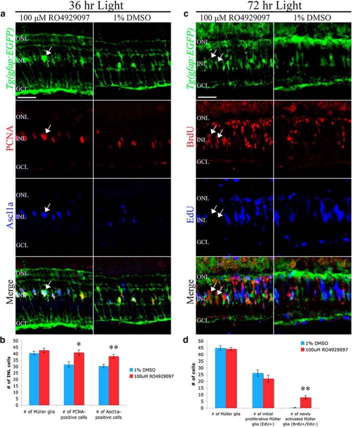
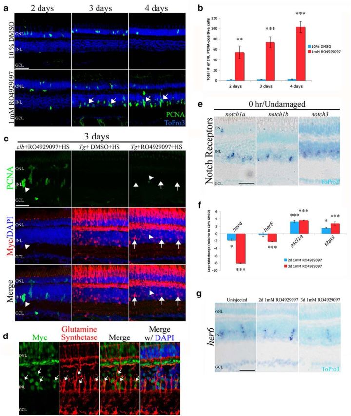
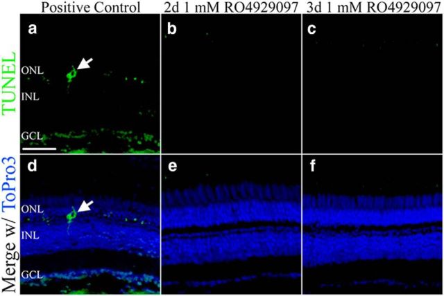
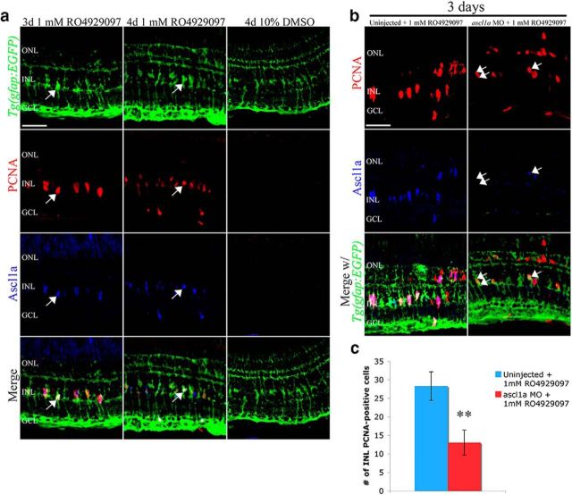
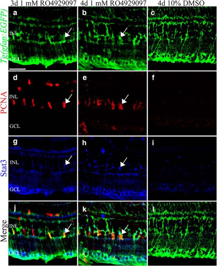
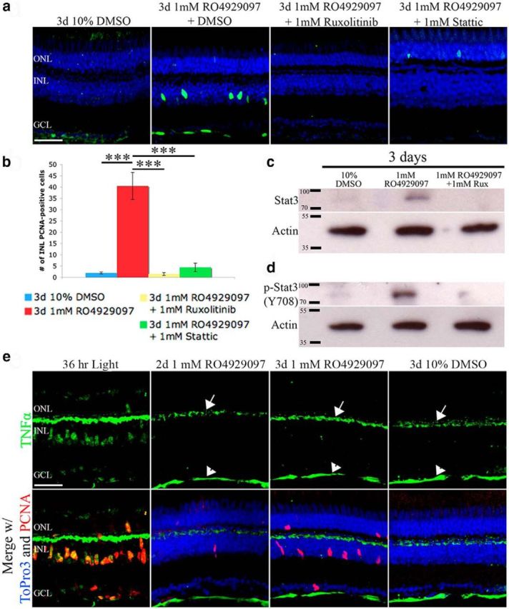
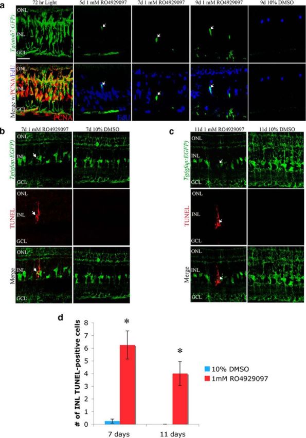
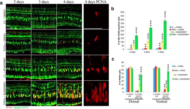
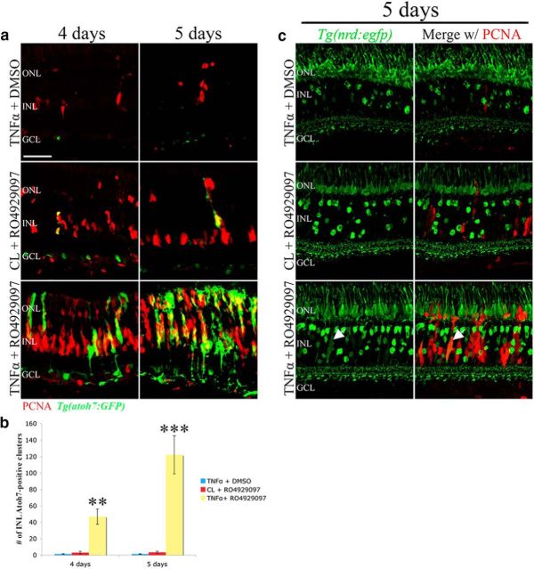
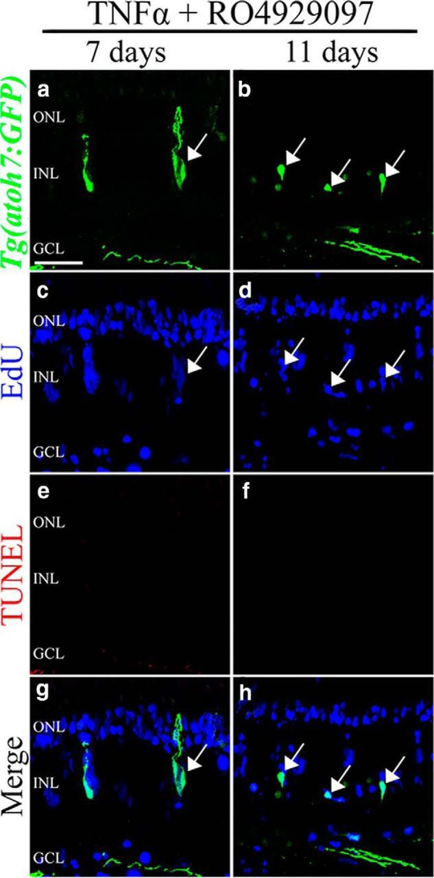
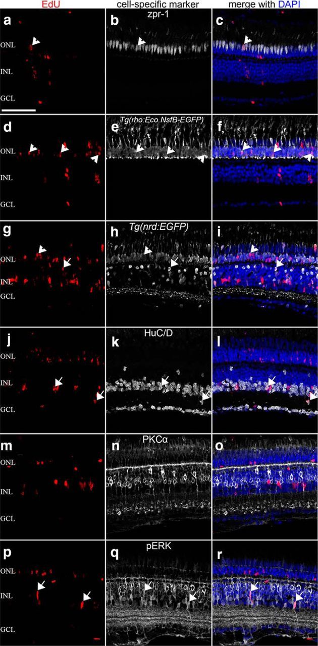
Retinal damage in teleosts, unlike mammals, induces robust Müller glia-mediated regeneration of lost neurons. We examined whether Notch signaling regulates Müller glia proliferation in the adult zebrafish retina and demonstrated that Notch signaling maintains Müller glia in a quiescent state in the undamaged retina. Repressing Notch signaling, through injection of the γ-secretase inhibitor RO4929097, stimulates a subset of Müller glia to reenter the cell cycle without retinal damage. This RO4929097-induced Müller glia proliferation is mediated by repressing Notch signaling because inducible expression of the Notch Intracellular Domain (NICD) can reverse the effect. This RO4929097-induced proliferation requires Ascl1a expression and Jak1-mediated Stat3 phosphorylation/activation, analogous to the light-damaged retina. Moreover, coinjecting RO4929097 and TNFα, a previously identified damage signal, induced the majority of Müller glia to reenter the cell cycle and produced proliferating neuronal progenitor cells that committed to a neuronal lineage in the undamaged retina. This demonstrates that repressing Notch signaling and activating TNFα signaling are sufficient to induce Müller glia proliferation that generates neuronal progenitor cells that differentiate into retinal neurons, mimicking the responses observed in the regenerating retina.

Keywords: Ascl1; Müller glia; Notch signaling; Stat3; quiescence; retinal regeneration.


Figures

Figure 1.
Figure 1.
Notch-inhibited retinas exhibit increased Müller glia activation and proliferation after light damage. a, Adult Tg(gfap:EGFP)nt11 zebrafish (green, row 1) were light-damaged for 24 h and then intravitreally injected with either 100 μm RO4929097 or 1% DMSO. After 36 h of constant light, RO4929097-treated retinas (column 1) exhibited increased numbers of PCNA-positive (red, row 2) and Ascl1a-positive (blue, row 3) Müller glia relative to DMSO-treated retinas (column 2). Arrows (column 1) indicate Müller glia nuclei double-positive for Ascl1a and PCNA. b, Histogram depicting no change in the number of INL Müller glia but a significant increase in the number of PCNA-positive and Ascl1a-positive INL cells in the RO4929097-treated light-damaged Tg(gfap:EGFP)nt11 retinas (n = 8). c, Adult Tg(gfap:EGFP)nt11 zebrafish (green, row 1) were intraperitoneally injected with EdU at 36 h of constant light, intravitreally injected with either 100 μm RO4929097 (column 1) or 1% DMSO (column 2) at 48 and 60 h, pulsed intraperitoneally with BrdU at 63 and 66 h, and immunolabeled at 72 h of light for EGFP (green, row 1), BrdU (red, row 2), and EdU (blue, row 3). The RO4929097-treated retinas (column 1) contained increased numbers of BrdU-positive INL cell clusters that were EdU-negative (arrows) relative to the DMSO control (column 2). d, Histogram depicting no change in either the number of EGFP-positive or EdU-positive Müller glia at 72 h of light-damage in DMSO or RO492097-treated retinas. The number of newly activated BrdU-positive/EdU-negative cell clusters, however, were significantly increased by 72 h in RO492097-treated retinas relative to the DMSO control retinas (n = 8). GCL, Ganglion cell layer; INL, inner nuclear layer; ONL, outer nuclear layer; PCNA, proliferating cellular nuclear antigen. Scale bars, 25 μm. Student's t test, *p < 0.05. **p < 0.01.
Figure 2.
Figure 2.
Inhibition of Notch signaling induces Müller glia proliferation in the absence of retinal damage. a, Intraperitoneal injection of 1 mm RO4929097 (bottom row) into AB zebrafish demonstrates an increased number of PCNA-positive INL cells (green, arrows) relative to the 1% DMSO-injected controls (top row). b, RO4929097 treatment results in a significant increase in the number of PCNA-positive INL cells relative to the DMSO control at 2, 3, and 4 d (n ≥ 7). c, Adult albino zebrafish were intraperitoneally injected with RO4929097 (alb+RO4929097+HS, column 1), and Tg(hsp70l:Gal4);Tg(UAS:myc-notch1a-intra) transgenic zebrafish were intraperitoneally injected with either RO4929097 (Tg+RO4929097+HS, column 3) or DMSO (Tg+DMSO+HS, column 2) and heat-shocked for 3 d. NICD-Myc (red) was expressed in the heat-shocked Tg(hsp70l:Gal4);Tg(UAS:myc-notch1a-intra) zebrafish and not in the heat-shocked albino fish. Expression of NICD-Myc reduced the number of PCNA-positive INL cells (green) that were induced by RO4929097. NICD-Myc-positive Müller glia (arrows) did not express PCNA. d, Tg(hsp70l:Gal4);Tg(UAS:myc-notch1a-intra) zebrafish were intraperitoneally injected with RO4929097 and heat-shocked for 3 d. The NICD-Myc fusion protein was induced in the glutamine synthetase-positive Müller glia (arrows). e, notch1a, notch1b, and notch3 mRNA expression was localized to the basal INL of the undamaged retina by in situ hybridization. f, qRT-PCR revealed that RO4929097 inhibition of Notch signaling significantly reduced the transcript levels of the Notch-specific targets her4 and her6 at 2 and 3 d and increased expression of the activated Müller glia markers ascl1a and stat3 (n = 4). g, Tissue section in situ hybridization verified a downregulation of the Notch downstream target, her6, by 3 d of RO4929097 treatment. GCL, Ganglion cell layer; INL, inner nuclear layer; ONL, outer nuclear layer; PCNA, proliferating cellular nuclear antigen. Scale bars, 25 μm. ToPro3 (a, e, g) and DAPI staining (c, d) were used to distinguish the three nuclear layers. *p < 0.05 (Student's t test). **p < 0.01 (Student's t test). ***p < 0.001 (Student's t test).
Figure 3.
Figure 3.
Cell death is not increased during RO4929097-induced inhibition of Notch signaling. a, d, Adult albino zebrafish that were treated with constant light for 16 h displayed TUNEL-positive cells (green, arrow) in the retina. Sections were counterstained with ToPro3 (blue) to label nuclei. b, c, e, f, Adult AB zebrafish that were intraperitoneally injected with 1 mm RO4929097 for either 2 d (b, e) or 3 d (c, f) exhibited no TUNEL-positive cells in the ONL, INL, or GCL. GCL, Ganglion cell layer; INL, inner nuclear layer; ONL, outer nuclear layer. Scale bars, 25 μm.
Figure 4.
Figure 4.
Inhibiting Notch signaling in the undamaged retina induces Ascl1a-dependent Müller glia proliferation. a, Undamaged adult Tg(gfap:EGFP)nt11 zebrafish (green, row 1) were intraperitoneally injected with either 1 mm RO4929097 or 10% DMSO. Confocal images of 1 mm RO4929097-treated retinas immunolabeled for PCNA (red, row 2) or Ascl1a (blue, row 3) at 3 and 4 d (columns 1 and 2, respectively) show increased numbers of both PCNA-positive and Ascl1a-positive Müller glia (merge) relative to the DMSO-injected controls (column 3). Additionally, the PCNA-positive cells colabeled (arrow) with the Ascl1a-positive activated Müller glia. b, Tg(gfap:EGFP)nt11 were either intraperitoneally injected with 1 mm RO4929097 (column 1) or intravitreally injected and electroporated with ascl1a MO (column 2) followed by intraperitoneal injection with 1 mm RO4929097 for 3 d. Knockdown of Ascl1a expression decreased the number of PCNA-positive (red, row 1) and Ascl1a-expressing (blue row 2) cells. Although Ascl1a levels were clearly decreased in the morphant retina, a very small number of Ascl1a-positive (single arrow) and Ascl1a-negative (double arrow) PCNA-expressing cells remained. c, Histogram depicting a significant decrease in the number of RO4929097-induced PCNA-positive cells in the INL following ascl1a morpholino knockdown (n = 8). GCL, Ganglion cell layer; INL, inner nuclear layer; ONL, outer nuclear layer; PCNA, proliferating cellular nuclear antigen; MO, morpholino. Scale bars, 25 μm. **p < 0.01 (Student's t test).
Figure 5.
Figure 5.
Inhibiting Notch signaling in the undamaged retina induces Stat3 expression in proliferating Müller glia. Undamaged Tg(gfap:EGFP)nt11 zebrafish were intraperitoneally injected with either 1 mm RO4929097 or 10% DMSO for 3 or 4 d and immunolabeled for EGFP (a–c, green), PCNA (d–f, red), and Stat3 (g–i, blue). 1 mm RO4929097-treated Tg(gfap:EGFP)nt11 retinas at 3 d (column 1) and 4 d (column 2) showed increased numbers of PCNA-positive (d, e, red) and Stat3-positive (g, h, blue) cells that colabeled EGFP-positive Müller glia (j, k, l, merge arrows) relative to the DMSO-injected controls (column 3). GCL, Ganglion cell layer; INL, inner nuclear layer; ONL, outer nuclear layer; PCNA, proliferating cellular nuclear antigen. Scale bars, 25 μm.
Figure 6.
Figure 6.
Jak-mediated Stat3 phosphorylation/activation is necessary for the Notch-inhibited undamaged retina to proliferate independent of TNFα. a, albino fish were intraperitoneally injected with DMSO, RO4929097 and DMSO, RO4929097 and ruxolitinib, or RO4929097 and Stattic for 3 d. Whereas RO4929097 and DMSO was sufficient to induce PCNA-positive INL cells, blocking the Stat3 signaling pathway with either ruxolitinib or Stattic inhibited the RO4929097-induced proliferation. b, Histogram depicting that the number of PCNA-positive INL cells in RO4929097-treated undamaged retinas was significantly reduced by cotreating with either ruxolitinib or Stattic (n = 8). c, d, Total protein lysates from DMSO, RO4929097 and DMSO, RO4929097 and ruxolitinib-treated retinas confirmed increased total Stat3 (c) and p-Stat3–708 (d) protein levels in the RO4929097-treated undamaged retinas and near control levels in the RO4929097 and ruxolitinib-cotreated retinas. β-Actin was used as a loading control. e, Confocal images of 36 h light-damaged retina (column 1) that was immunolabeled for TNFα (green) and PCNA (red) show robust TNFα expression in PCNA-positive INL cells. Undamaged albino zebrafish were intraperitoneally injected with either 1 mm RO4929097 or 10% DMSO vehicle control for either 2 or 3 d (columns 2, 3, and 4). The RO4929097 and DMSO control retinal sections were immunolabeled for TNFα (green, row 1) and merged with PCNA (red, row 2) and the nuclear marker ToPro3 (blue, row 2). Although TNFα expression was detected in both the outer plexiform layer and nerve fiber layer (arrow and arrowhead, respectively), there was no detectable TNFα expression in the INL or ONL of either the RO4929097 or DMSO-treated retinas. This demonstrates that RO4929097-induced PCNA expression in the INL is not the result of increased TNFα expression in the INL Müller glia. GCL, Ganglion cell layer; INL, inner nuclear layer; ONL, outer nuclear layer; PCNA, proliferating cellular nuclear antigen; Rux, ruxolitinib. Scale bars, 25 μm. ***p < 0.001 (one-way ANOVA and Tukey's post hoc test).
Figure 7.
Figure 7.
The proliferating neuronal progenitors produced by inhibiting Notch signaling in the undamaged retina do not differentiate and undergo cell death. Undamaged albino Tg(atoh7:GFP) zebrafish (a) and albino Tg[gfap:EGFP]nt11 transgenic zebrafish (b, c) were intraperitoneally injected with either 1 mm RO4929097 or 10% DMSO every 12 h for 4 d, EdU was coinjected at 1.5, 2.5, and 3.5 d, and retinas were collected at 5, 7, 9, or 11 d. a, Confocal image of Tg(atoh7:GFP) (green, column 1) retinas that were light-damaged for 72 h displayed robust PCNA expression (red, column 1) in the GFP-positive INL clusters. RO4929097-treated Tg(atoh7:GFP) zebrafish (columns 2–4) displayed large numbers of EdU-positive INL cells (blue, lower row) relative to the DMSO control (column 5). Although limited, there was also an increase in the number of GFP-positive INL cells that colabel with EdU-positive INL cells (arrow) in the RO4929097-treated retinas relative to the DMSO-treated retinas. b, c, Confocal images of Tg[gfap:EGFP]nt11 transgenic zebrafish (which express EGFP in the Müller glia from the gfap promoter) treated with either 1 mm RO4929097 (column 1) or 10% DMSO (column 2) for 7 d (b) and 11 d (c) and labeled with anti-GFP to identify the Müller glia (green, row 1) and TUNEL (red, row 2). Arrows indicate Müller glia that are TUNEL-positive. d, Histogram depicting an increased number of TUNEL-positive INL cells at both 7 and 11 d in RO4929097-injected retinas compared with 10% DMSO (n = 5). GCL, Ganglion cell layer; INL, inner nuclear layer; ONL, outer nuclear layer; PCNA, proliferating cellular nuclear antigen. Scale bars, 25 μm. Student's t test, *p < 0.05.
Figure 8.
Figure 8.
Inhibition of Notch signaling in combination with exogenous TNFα act synergistically to induce robust Müller glia proliferation. a, Adult Tg(gfap:EGFP)nt11 transgenic zebrafish were intraperitoneally injected with either 1 mm RO4929097 or 10% DMSO in combination with an intravitreal injection of either purified recombinant TNFα or purified control bacterial lysate (CL) for 2–4 d. The TNFα/DMSO, RO4929097/CL, and TNFα/RO4929097 coinjected retinas possessed signifcantly more PCNA-positive cells (red) at 2, 3, and 4 d of injections relative to the CL/DMSO coinjected control. The fourth column (4 d PCNA) contains higher-magnification images of PCNA labeling to highlight the single INL cells in the TNFα/DMSO coinjected retina, the single and double cells in the RO4929097/CL coinjected retina, and the columns containing several PCNA-positive cells in the TNFα/RO4929097 coinjected retina. b, Histogram depicting a significant increase in the number of PCNA-positive INL cells in the TNFα/DMSO, RO4929097/CL, and RO4929097/TNFα-coinjected retinas compared with the CL/DMSO coinjected control (n ≥ 6). c, Histogram showing a significant increase in the number of dorsal and ventral PCNA-positive Müller glia in the TNFα/DMSO, RO4929097/CL, and RO4929097/TNFα-coinjected retinas compared with the CL/DMSO coinjected control (n ≥ 6), whereas the total number of Müller glia remained unchanged in all four treatment groups. GCL, Ganglion cell layer; INL, inner nuclear layer; ONL, outer nuclear layer; PCNA, proliferating cellular nuclear antigen. Scale bars, 25 μm. **p < 0.01 (two-way ANOVA and Tukey's post hoc test). ***p < 0.001 (two-way ANOVA and Tukey's post hoc test).
Figure 9.
Figure 9.
Robust proliferation stimulated by coinjection of RO4929097 and TNFα stimulates neuronal progenitors to commit to a neuronal fate. Tg(atoh7:GFP) or Tg(nrd:GFP) zebrafish were coinjected with TNFα/DMSO, RO4929097/CL, and RO4929097/TNFα for 4 d. a, The Tg(atoh7:GFP) zebrafish were collected at 4 and 5 d and immunolabeled for PCNA (red). The RO4929097/TNFα coinjected retinas had significantly more GFP-positive cell clusters that robustly colabeled with PCNA than either of the other two treatments. b, Histogram depicting the increased number of GFP-positive INL clusters present in the RO4929097/TNFα cotreated retinas relative to the other two treatments (n = 7). c, The Tg(nrd:egfp) zebrafish were collected at 5 d and immunolabeled for PCNA (red). The RO4929097/TNFα coinjected retinas had more diffuse GFP-positive cells (representing committed neuronal progenitor cells) that robustly colabeled with PCNA (arrows) than either of the other two treatments. GCL, Ganglion cell layer; INL, inner nuclear layer; ONL, outer nuclear layer; PCNA, proliferating cellular nuclear antigen. Scale bars, 25 μm. **p < 0.01 (two-way ANOVA and Tukey's post hoc test). ***p < 0.001 (two-way ANOVA and Tukey's post hoc test).
Figure 10.
Figure 10.
Coinjection of RO4929097 and TNFα reduces neuronal progenitor cell death. Tg(atoh7:GFP) zebrafish were coinjected with TNFα and RO4929097 and labeled with EdU. After 7 d (a, c, e, g) and 11 d (b, d, f, h), the retinas were immunolabeled for GFP (a, b, g, h), EdU (c, d, g, h), and TUNEL (e–h). The merged images of the three different labels are shown (g, h). After 7 d, the retina possessed EdU-labeled cells that were GFP-positive (arrows). No TUNEL-positive cells were detected in the retinas at either day. GCL, Ganglion cell layer; INL, inner nuclear layer; ONL, outer nuclear layer. Scale bars, 25 μm.
Figure 11.
Figure 11.
Coinjection of RO4929097 and TNFα is sufficient to induce neuronal progenitor cells to differentiate into most types of retinal neurons. Wild-type, Tg(nrd:egfp), or Tg(rho:Eco.NfsB-EGFP)nt19 zebrafish were coinjected with TNFα and RO4929097 and labeled with EdU. After 11 d, the retinas were immunolabeled for EdU (a, c, d, f, g, i, j, l, m, o, p, r), Zpr-1 (b, c), EGFP (e, f, h, i), HuC/D (k, l), PKCα (n, o), pERK (q, r), and DAPI (c, f, i, l, o, r). EdU-labeled cone (a–c) and rod (d–i) are marked with arrowheads. EdU-labeled bipolar cells (g–i), amacrine cells (j–l), and Müller glia (p–r) are indicated with arrows. There are no detectable EdU-labeled ganglion cells (j–l) or on-bipolar cells (m–o). GCL, Ganglion cell layer; INL, inner nuclear layer; ONL, outer nuclear layer. Scale bars, 25 μm.

References

    1. Bailey TJ, Fossum SL, Fimbel SM, Montgomery JE, Hyde DR. The inhibitor of phagocytosis, O-phospho-L-serine, suppresses Müller glia proliferation and cone cell regeneration in the light-damaged zebrafish retina. Exp Eye Res. 2010;91:601–612. doi: 10.1016/j.exer.2010.07.017. - DOI - PMC - PubMed
    1. Bernardos RL, Lentz SI, Wolfe MS, Raymond PA. Notch-Delta signaling is required for spatial patterning and Müller glia differentiation in the zebrafish retina. Dev Biol. 2005;278:381–395. doi: 10.1016/j.ydbio.2004.11.018. - DOI - PubMed
    1. Bhattacharya S, Das AV, Mallya KB, Ahmad I. Ciliary neurotrophic factor-mediated signaling regulates neuronal versus glial differentiation of retinal stem cells/progenitors by concentration-dependent recruitment of mitogen-activated protein kinase and Janus kinase-signal transducer and activator of transcription pathways in conjunction with Notch signaling. Stem Cells. 2008;26:2611–2624. doi: 10.1634/stemcells.2008-0222. - DOI - PubMed
    1. Bringmann A, Pannicke T, Grosche J, Francke M, Wiedemann P, Skatchkov SN, Osborne NN, Reichenbach A. Müller cells in the healthy and diseased retina. Prog Retin Eye Res. 2006;25:397–424. doi: 10.1016/j.preteyeres.2006.05.003. - DOI - PubMed
    1. Chapouton P, Skupien P, Hesl B, Coolen M, Moore JC, Madelaine R, Kremmer E, Faus-Kessler T, Blader P, Lawson ND, Bally-Cuif L. Notch activity levels control the balance between quiescence and recruitment of adult neural stem cells. J Neurosci. 2010;30:7961–7974. doi: 10.1523/JNEUROSCI.6170-09.2010. - DOI - PMC - PubMed

Publication types

MeSH terms

Substances

LinkOut - more resources