. 2014 Oct 22;34(43):14430-42.
doi: 10.1523/JNEUROSCI.5366-13.2014.

Adult neurogenesis restores dopaminergic neuronal loss in the olfactory bulb

Affiliations

Adult neurogenesis restores dopaminergic neuronal loss in the olfactory bulb

Françoise Lazarini et al. J Neurosci. .

Abstract

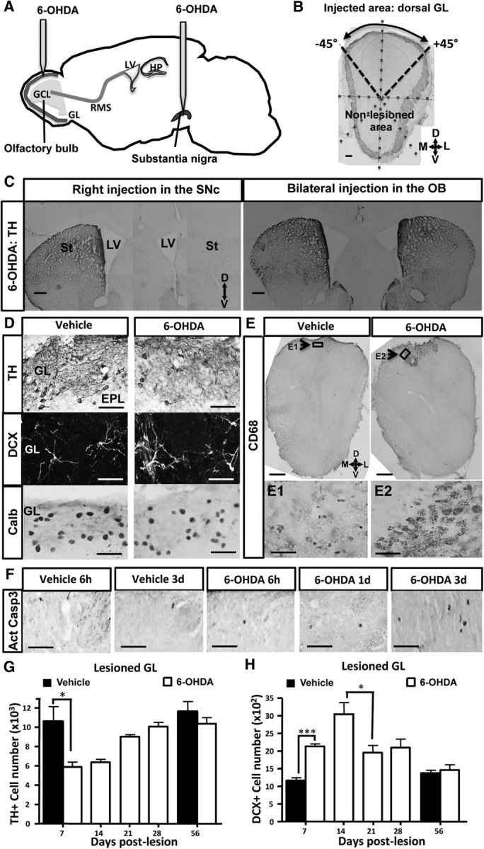
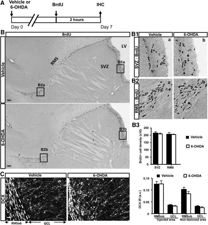
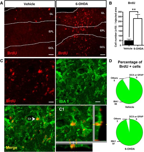
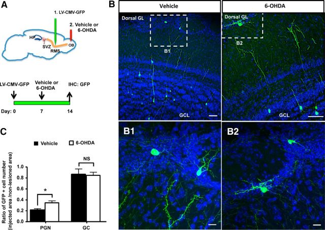
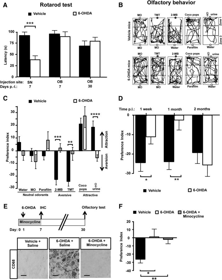
Subventricular zone (SVZ) neurogenesis continuously provides new GABA- and dopamine (DA)-containing interneurons for the olfactory bulb (OB) in most adult mammals. DAergic interneurons are located in the glomerular layer (GL) where they participate in the processing of sensory inputs. To examine whether adult neurogenesis might contribute to regeneration after circuit injury in mice, we induce DAergic neuronal loss by injecting 6-hydroxydopamine (6-OHDA) in the dorsal GL or in the right substantia nigra pars compacta. We found that a 6-OHDA treatment of the OB produces olfactory deficits and local inflammation and partially decreases the number of neurons expressing the enzyme tyrosine hydroxylase (TH) near the injected site. Blockade of inflammation by minocycline treatment immediately after the 6-OHDA administration rescued neither TH(+) interneuron number nor the olfactory deficits, suggesting that the olfactory impairments are most likely linked to TH(+) cell death and not to microglial activation. TH(+) interneuron number was restored 1 month later. This rescue resulted at least in part from enhanced recruitment of immature neurons targeting the lesioned GL area. Seven days after 6-OHDA lesion in the OB, we found that the integration of lentivirus-labeled adult-born neurons was biased: newly formed neurons were preferentially incorporated into glomerular circuits of the lesioned area. Behavioral rehabilitation occurs 2 months after lesion. This study establishes a new model into which loss of DAergic cells could be compensated by recruiting newly formed neurons. We propose that adult neurogenesis not only replenishes the population of DAergic bulbar neurons but that it also restores olfactory sensory processing.

Keywords: Parkinson's disease; adult neural stem cell; innate responses; interneuron; microglia; regenerative medicine.


Figures

Figure 1.
Figure 1.
Transient dopamine cell loss after a 6-OHDA-induced lesion of the OB. A, Design of the experimental procedure. A 6-OHDA-containing solution, or vehicle alone, was infused into the right SN or into the dorsal part of each OB. B, TH immunostaining delineates the injected and noninjected areas in a coronal OB section. The image was divided into 8 sectors of 45°. C, TH immunoreactivity of coronal sections of striata and SVZ at 1 month after 6-OHDA injection into the right SN (left) or the two OBs (right). The efficacy of DA denervation is demonstrated by low levels of TH expression in the right striatum and right SVZ after neurotoxin injection into the right SN (left), depicting the extent and specificity of the lesion. Conversely, the right panel represents no alteration of TH expression in the two striata and SVZ after injection of 6-OHDA into the OB. D, Immunoreactivity for TH (top), DCX (middle), and Calb (bottom) of the lesioned GL at 7 d after injection into the OB of vehicle (left) or 6-OHDA (right). 6-OHDA reduces the number of DAergic cells expressing TH in the dorsal GL. Conversely, 6-OHDA increases DCX number but does not change Calb number in the same area. E, Immunoreactivity for CD68 (E1, E2, top) of the dorsal OB at 7 d after injection into the OB of vehicle (left) or 6-OHDA (right). 6-OHDA activates microglial cells as revealed by CD68 staining. Note the restricted presence of activated microglia in the dorsal part of the OB, depicting the extent and specificity of the lesion. Arrow indicates the area shown at higher magnification in E1 and E2. F, Time course of apoptotic cells expressing active cleaved casp 3 in the lesioned GL after 6 h, 1 d, and 3 d after 6-OHDA injection. The number of apoptotic cells is increased at day 3 after OB lesion. G, Time course of TH+ PGNs located in the lesioned GL after OB lesion (n = 4 or 5 animals per group). The number of TH-expressing neurons is transiently reduced in the dorsal GL after OB lesion. H, Time course of DCX-positive PGNs located in the lesioned GL after OB lesion (n = 4 or 5 per group). The number of developing neurons expressing DCX peaks at day 14 in the dorsal GL after OB lesion and resumes 21 d later. *p < 0.05. ***p < 0.001. Scale bars: B, C, 100 μm; D, 20 μm; E, 100 μm; E1, E2, 20 μm.
Figure 2.
Figure 2.
6-OHDA-induced lesion of the OB spares the first steps of neurogenesis. A, Design of the experiments. Vehicle, or 6-OHDA, was bilaterally injected into the GL, and mice were perfused 7 d later. To label proliferating cells, BrdU was injected 2 h before perfusion. B, Sagittal sections of the forebrain injected with vehicle (top) or 6-OHDA (bottom) showing the different neurogenic zones labeled with BrdU. B1, B2, Higher magnifications of areas indicated in the sections. B3, BrdU-positive cell density in the SVZ and RMS at day 7 after lesioning. C, Coronal sections of the OB of vehicle (left) or 6-OHDA (middle) showing the RMSob and the GCL labeled with DCX. Right, DCX immunoreactivity (IR, in arbitrary units, a.u.) in the RMSob and GCL of injected and nonlesioned areas quantified 7 d after lesion. Scale bars: B, 250 μm; C, 25 μm.
Figure 3.
Figure 3.
Microglial cells, but not neuroblasts, proliferate in the 6-OHDA-lesioned OB. A, BrdU immunostaining in the OB of a vehicle (left) and a 6-OHDA-injected OB (right) at 7 d after lesion. BrdU-positive cells were restricted to the dorsal part of the OB, mainly in the GL, but also in the external plexiform layer and GCL. B, Quantification of BrdU-positive cells in the injected area (dorsal part of the OB) at 7 d after lesion (n = 4 mice per group). C, BrdU and IBA1 immunostaining in the GL of a 6-OHDA-injected OB at 7 d after lesion. BrdU was only found in microglial cells expressing IBA1. Arrow indicates the cell shown at higher magnification in C1. D, Percentage of BrdU-positive cells colabeled with one of three cell markers: GFAP for astrocytes, DCX for developing neurons, and IBA1 for active microglial cells. Cell counts were performed in the dorsal GL of vehicle and 6-OHDA-lesioned mice at 7 d after lesion (n = 4 mice per group, ∼100 cells analyzed per mouse). Nearly all fast-proliferating cells positive for BrdU coexpressed IBA1. **p < 0.01, compared with vehicle. Scale bars: A, 50 μm; C, 10 μm.
Figure 4.
Figure 4.
Preferential incorporation of lentivirus-labeled adult-born PGNs in the local area of DAergic neuron ablation. A, Overview of the experimental procedure. Mice were bilaterally injected into the RMS by a replication-defective lentivirus expressing eGFP to label migrating newborn cells at the time of injection. Seven days later, vehicle, or 6-OHDA, was bilaterally injected into the GL, and mice were analyzed 2 weeks later. B, GFP staining in the dorsal OB at 2 weeks after viral injection in vehicle and 6-OHDA-lesioned mice. B1, B2, Delineated areas are shown at higher magnification. C, Ratio of the number of virus-labeled new neurons in the injected area over the one counted in the noninjected area (n = 4 mice per group). This ratio is more elevated for PGN in 6-OHDA-injected mice, indicating a bias for incorporating immature neurons into the lesioned area. *p < 0.05, compared with vehicle. NS, Not significant. Scale bars: B1, B2, 20 μm.
Figure 5.
Figure 5.
Behavioral consequences of challenging bulbar DAergic interneurons. A, Mean latency before falling down for the rotarod test are shown for mice injected with vehicle or 6-OHDA into the right SN or the two OBs at day 7 and day 30 after lesion (n = 8–23 per group). Mice injected into the SN exhibited shorter latencies compared with the control groups (i.e., vehicle-injected mice and mice treated with bilateral injection of 6-OHDA into the OB), indicating motor deficits. B, Representative tracing of mouse locomotion at 7 d after lesion, during a 3 min exposure to the tested odorant and to its vehicle. Panels represent the third stage of one session of the innate olfactory test habituation: exposure to the tested odorant in zone 1 and to its vehicle in zone 3. Top, Tracings of a vehicle-injected mouse (“vehicle mice”). Bottom, Tracings of a 6-OHDA-injected mouse (“6-OHDA mice”). The position of the mouse was quantified in the cage test by measuring the time spent in zone 1 and zone 3. The tested odorants were MO, TMT, 2-MB acid, Coco Pop cereals, and pool of female urines (“female urine”). Vehicle odorants were MO, water, or Parafilm as appropriate. Odorants were positioned in the middle of zone 1, except for Coco Pop cereals, which were exposed in the left part of zone 1. Whereas vehicle mice avoid aversive olfactory cues (TMT and 2-MB acid), 6-OHDA-treated mice showed no interest in these odorants. Moreover, whereas female urine attracted the vehicle mice, the 6-OHDA mice showed no interest to this social scent. C, Preference index values for mice injected into the two OBs by vehicle (black columns) or 6-OHDA (white columns) at 7 d after lesion (n = 14–20 mice per group, per odorants). Olfactory cues were chosen to be neutral (water, MO, Parafilm), aversive (2-MB acid, TMT), or attractive (Coco Pop cereals, female urine). Preference index = time spent in zone 1 during odorant trial − time spent in zone 1 during vehicle trial. Positive values indicate attraction and negative value repulsion. Female urine was strongly attractive for vehicle male mice but not for 6-OHDA-injected mice. 2-MB acid and TMT elicited robust aversion for vehicle mice, but not for 6-OHDA-injected mice. D, Preference index values for mice injected into the two OBs by vehicle (black) or 6-OHDA (white) at 1 week, 1 month, and 2 months after lesion (n = 15–18 mice per group). The tested odorant is the aversive odorant TMT. Whereas TMT induces a strong avoidance for vehicle mice, the response to this olfactory cue was altered in 6-OHDA-treated mice but restored at 2 months after lesion. E, Minocycline treatment blocks the strong neuroinflammation induced by 6-OHDA lesion. Top, Design of the experimental procedure. Minocycline or saline was administered during 8 d, starting 1 d before 6-OHDA lesion of the OB. A 6-OHDA-containing solution, or vehicle alone, was infused into the dorsal part of each OB. Bottom, CD68 staining in the dorsal lesioned OB at 1 week after 6-OHDA injection in vehicle (left), 6-OHDA-lesioned (center), and minocycline-treated lesioned (right) mice. F, Preference index values for mice treated with saline or minocycline (gray) for 1 week and injected at day 1 into the two OBs by vehicle (black) or 6-OHDA (white) at 1 month after lesion (n = 7–10 mice per group). The response to TMT is unchanged by minocycline treatment. *p < 0.05. **p < 0.01. ***p < 0.001. ****p < 0.0001. Scale bar, 10 μm.

Comment in

References

    1. Adam Y, Mizrahi A. Circuit formation and maintenance: perspectives from the mammalian olfactory bulb. Curr Opin Neurobiol. 2010;20:134–140. doi: 10.1016/j.conb.2009.11.001. - DOI - PubMed
    1. Adam Y, Mizrahi A. Long-term imaging reveals dynamic changes in the neuronal composition of the glomerular layer. J Neurosci. 2011;31:7967–7973. doi: 10.1523/JNEUROSCI.0782-11.2011. - DOI - PMC - PubMed
    1. Akiyama H, McGeer PL. Microglial response to 6-hydroxydopamine-induced substantia nigra lesions. Brain Res. 1989;489:247–253. doi: 10.1016/0006-8993(89)90857-3. - DOI - PubMed
    1. Alonso M, Lepousez G, Sebastien W, Bardy C, Gabellec MM, Torquet N, Lledo PM. Activation of adult-born neurons facilitates learning and memory. Nat Neurosci. 2012;15:897–904. doi: 10.1038/nn.3108. - DOI - PubMed
    1. Alvarez-Buylla A, Lim DA. For the long run: maintaining germinal niches in the adult brain. Neuron. 2004;41:683–686. doi: 10.1016/S0896-6273(04)00111-4. - DOI - PubMed

Publication types