. 2015 Jan;352(1):148-55.
doi: 10.1124/jpet.114.217976. Epub 2014 Oct 22.

Evidence for α-helices in the large intracellular domain mediating modulation of the α1-glycine receptor by ethanol and Gβγ

Affiliations

Evidence for α-helices in the large intracellular domain mediating modulation of the α1-glycine receptor by ethanol and Gβγ

Carlos F Burgos et al. J Pharmacol Exp Ther. 2015 Jan.

Erratum in

  • J Pharmacol Exp Ther. 2015 Feb;352(2):325

Abstract

The α1-subunit containing glycine receptors (GlyRs) is potentiated by ethanol, in part, by intracellular Gβγ actions. Previous studies have suggested that molecular requirements in the large intracellular domain are involved; however, the lack of structural data about this region has made it difficult to describe a detailed mechanism. Using circular dichroism and molecular modeling, we generated a full model of the α1-GlyR, which includes the large intracellular domain and provides new information on structural requirements for allosteric modulation by ethanol and Gβγ. The data strongly suggest the existence of an α-helical conformation in the regions near transmembrane (TM)-3 and TM4 of the large intracellular domain. The secondary structure in the N-terminal region of the large intracellular domain near TM3 appeared critical for ethanol action, and this was tested using the homologous domain of the γ2-subunit of the GABAA receptor predicted to have little helical conformation. This region of γ2 was able to bind Gβγ and form a functional channel when combined with α1-GlyR, but it was not sensitive to ethanol. Mutations in the N- and C-terminal regions introduced to replace corresponding amino acids of the α1-GlyR sequence restored the ability to be modulated by ethanol and Gβγ. Recovery of the sensitivity to ethanol was associated with the existence of a helical conformation similar to α1-GlyR, thus being an essential secondary structural requirement for GlyR modulation by ethanol and G protein.


Figures

Fig. 1.
Fig. 1.
Full model of the α1-GlyR subunit. (A) Representation of the α1 monomer including the large intracellular domain determined by ab initio technique. The other regions were modeled using the template from the GLUC crystallographic data (PDB ID 3RIF). Two helical structures are present in the LIL, and basic amino acids described as important for modulation of GlyR by ethanol and Gβγ are indicated for reference (black arrows). The relative position of the membrane is shown by the dotted line. The rainbow colors represent the N- (blue) to C-(red) terminal progression of the protein. (B) Pentameric assembly of the GlyR composed of five identical α1-subunits. (Left) Side vision of the full channel highlighting the basic amino acid clusters 316–320 (magenta) located in the α-helix near TM3, and 385–386 (red) in the proximity to TM4 after the second helix predicted in the LIL C-terminal. (Right) Top view with subunits distinguished by different colors showing monomer distribution and pore formation; bottom view of the GlyR presenting the orientation of both helices and localization of the basic amino acid clusters (magenta and red). The segment between residues 334 and 370 corresponding to random conformation was removed.
Fig. 2.
Fig. 2.
Secondary structure of the LIL of the α1-subunit determined with circular dichroism. (A) Schematic representation of the LIL of α1-GlyR showing the full sequence and the three truncated proteins used. (B) CD spectra data from the LIL of α1 (green) and truncated proteins GlyR-NT (magenta), GlyR-IR (red), and GlyR-CT (blue) and the LIL of γ2 (gray) expressed as GST fusion proteins. (C) Secondary structure calculated from CD spectra of full α1-LIL, truncated proteins and γ2-LIL shown as a percentage of helix, β-sheet, and random conformation.
Fig. 3.
Fig. 3.
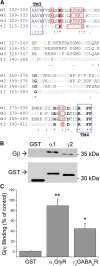
GST pull-down of the LIL of α1-GlyR, γ2-GABAAR and Gβγ. (A) Sequence alignments of the LILs of α-GlyRs and the γ2-GABAAR subunit. Bold letters show conserved residues and gray letters represent nonconserved amino acids. Basic residues near TM3 and TM4 are shown in red squares. (B) Western blot shows the interaction of α1-LIL GlyR and γ2-LIL GABAAR with Gβγ dimer by GST pull-down assays. Arrows indicate Gβ detected using a polyclonal anti-Gβ antibody and GST revealed by an anti-GST antibody, respectively. (C) Quantification of Gβ binding to the two LIL domains showing the relative amounts of bound Gβ normalized with their respective loaded GST fusion protein. *P < 0.05; **P < 0.01. Data are mean ± S.E.M., n = 4.
Fig. 4.
Fig. 4.
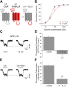
Characterization of the chimeric receptor α1-LIL-γ2 sensitivity to G protein and ethanol modulations. (A) Schematic representation shows the generation of the chimera α1-LIL-γ2. (B) Glycine concentration-response relationship for the α1-GlyR and the chimera α1-LIL-γ2. (C) Representative glycine-evoked current traces at the beginning (1 minute) and after 15 minutes of intracellular dialysis of the nonhydrolyzable analog GTPγS in HEK cells expressing the chimera α1-LIL-γ2. The concentration of glycine was 15 μM (EC10–20). (D) Effects of GTPγS on α1-GlyR and chimera α1-LIL-γ2 after 15 minutes of dialysis in terms of the percentage of potentiation of evoked currents in relation to the initial evoked current. (E) Representative glycine-evoked current traces (15 μM) in the absence and presence of 100 mM ethanol in α1-LIL-γ2. (F) Effects of 100 mM ethanol on α1-GlyR and chimera α1-LIL-γ2 in terms of percent of potentiation by 100 mM ethanol of 15 μM glycine-evoked currents. **P < 0.001. Data are mean ± S.E.M. (n = 6).
Fig. 5.
Fig. 5.
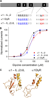
Characterization of chimeric receptor α1-LIL-γ2 ΔLIL containing homologous replacements of amino acids in regions near TM3 and -4. (A) Mutations in the chimera α1-LIL-γ2 to include the residues present in the α1-GlyR subunit. The α1-LIL-γ2 ΔLIL construct includes the cluster 309–313 RQHKE (blue) and the 385–386 basic cluster KK (red). (B) Glycine concentration-response curves for α1-GlyR, chimera α1-LIL-γ2 and modified chimera α1-LIL-γ2 ΔLIL. (C) Homology model of chimera α1-LIL-γ2 ΔLIL having a recovery of helical structure similar to that observed in the model of the α1-GlyR subunit.
Fig. 6.
Fig. 6.
Modulation of chimeric receptor α1-LIL-γ2 ΔLIL by Gβγ and ethanol. (A) Glycine-evoked current traces at the beginning (1 minute) and after 15 minutes of intracellular dialysis of the nonhydrolyzable analog GTPγS in HEK cells expressing the chimera α1-LIL-γ2 ΔLIL. The concentration of glycine was 15 μM (EC10–20). (B) Effects of GTPγS on chimera α1-LIL-γ2 and its modified version α1-LIL-γ2 ΔLIL after 15 minutes of dialysis as the percentage of potentiation of evoked currents with respect to the initial current amplitude. (C) Glycine-evoked current traces (15 μM) in the absence and presence of 100 mM ethanol recorded in HEK cells expressing the chimera α1-LIL-γ2 ΔLIL. (D) Effects of 100 mM ethanol on chimera α1-LIL-γ2 and its modified version α1-LIL-γ2 ΔLIL shown as percent potentiation by 100 mM ethanol of 15 μM glycine-evoked currents. **P < 0.001. Data are mean ± S.E.M. from at least four cells.

References

    1. Aguayo LG, Pancetti FC. (1994) Ethanol modulation of the gamma-aminobutyric acidA- and glycine-activated Cl- current in cultured mouse neurons. J Pharmacol Exp Ther 270:61–69. - PubMed
    1. Aguayo LG, Tapia JC, Pancetti FC. (1996) Potentiation of the glycine-activated Cl- current by ethanol in cultured mouse spinal neurons. J Pharmacol Exp Ther 279:1116–1122. - PubMed
    1. Alexander NS, Preininger AM, Kaya AI, Stein RA, Hamm HE, Meiler J. (2014) Energetic analysis of the rhodopsin-G-protein complex links the α5 helix to GDP release. Nat Struct Mol Biol 21:56–63. - PMC - PubMed
    1. Bocquet N, Nury H, Baaden M, Le Poupon C, Changeux JP, Delarue M, Corringer PJ. (2009) X-ray structure of a pentameric ligand-gated ion channel in an apparently open conformation. Nature 457:111–114. - PubMed
    1. Bonacci TM, Ghosh M, Malik S, Smrcka AV. (2005) Regulatory interactions between the amino terminus of G-protein betagamma subunits and the catalytic domain of phospholipase Cbeta2. J Biol Chem 280:10174–10181. - PubMed

Publication types

MeSH terms

LinkOut - more resources