Review
. 2014 Oct 7:5:378.
doi: 10.3389/fphys.2014.00378. eCollection 2014.

CLC channel function and dysfunction in health and disease

Affiliations
Review

CLC channel function and dysfunction in health and disease

Gabriel Stölting et al. Front Physiol. .

Abstract

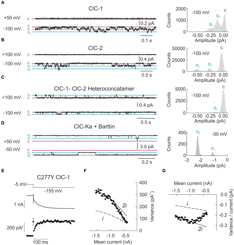
CLC channels and transporters are expressed in most tissues and fulfill diverse functions. There are four human CLC channels, ClC-1, ClC-2, ClC-Ka, and ClC-Kb, and five CLC transporters, ClC-3 through -7. Some of the CLC channels additionally associate with accessory subunits. Whereas barttin is mandatory for the functional expression of ClC-K, GlialCam is a facultative subunit of ClC-2 which modifies gating and thus increases the functional variability within the CLC family. Isoform-specific ion conduction and gating properties optimize distinct CLC channels for their cellular tasks. ClC-1 preferentially conducts at negative voltages, and the resulting inward rectification provides a large resting chloride conductance without interference with the muscle action potential. Exclusive opening at voltages negative to the chloride reversal potential allows for ClC-2 to regulate intracellular chloride concentrations. ClC-Ka and ClC-Kb are equally suited for inward and outward currents to support transcellular chloride fluxes. Every human CLC channel gene has been linked to a genetic disease, and studying these mutations has provided much information about the physiological roles and the molecular basis of CLC channel function. Mutations in the gene encoding ClC-1 cause myotonia congenita, a disease characterized by sarcolemmal hyperexcitability and muscle stiffness. Loss-of-function of ClC-Kb/barttin channels impairs NaCl resorption in the limb of Henle and causes hyponatriaemia, hypovolemia and hypotension in patients suffering from Bartter syndrome. Mutations in CLCN2 were found in patients with CNS disorders but the functional role of this isoform is still not understood. Recent links between ClC-1 and epilepsy and ClC-Ka and heart failure suggested novel cellular functions of these proteins. This review aims to survey the knowledge about physiological and pathophysiological functions of human CLC channels in the light of recent discoveries from biophysical, physiological, and genetic studies.

Keywords: Bartter syndrome; CLC channel; anion channel; leukencephalopathy; myotonia congenita; patch clamp.


Figures

Figure 1
Figure 1
The CLC family of transport proteins encompasses chloride channels and chloride/proton exchangers. (A) A phylogram demonstrates the early separation of the human CLC proteins into one branch of chloride channels encompassing ClC-1, ClC-2, ClC-Ka, and ClC-Kb. The chloride/proton antiporters encompass ClC-3 through −7. ClC-K channels require the subunit barttin while ClC-7 is dependent on the presence of Ostm1 for normal function. Recently, GlialCAM has been identified as accessory subunit of ClC-2. (B) A view of the dimer of cmClC [PDB ID: 3ORG (Feng et al., 2010)] in a ribbon presentation. One subunit is shown in light gray while the other subunit is shown in color ranging from the beginning of the B-helix in blue to the carboxy terminus in red. The upper part of the protein comprises the transmembrane core while the lower red part contains the carboxy termini with the CBS domains. (C) A closer view of some of the critical residues coordinating the chloride ions in ecClC (PDB ID: 1OTS). The magenta colored residues Y445 and S107 are responsible for the binding of chloride (green sphere) in Scen and are also proposed to be critical for slow gating of the CLC channels. E148 represents the so-called “gating glutamate” swinging from Scen to Sext and is thought to be tightly involved in fast protopore gating of CLC channels.
Figure 2
Figure 2
Whole-cell patch clamp recordings demonstrate the functional variability of human CLC channels. (A–D) Representative whole-cell current responses from HEK293T cells expressing ClC-1, ClC-2, ClC-Ka/barttin, or ClC-Kb/barttin to the indicated voltage protocols (left column). Right column shows the voltage dependences of the open probabilities of fast protopore (filled circles) and slow common gates (open circles) as well as of the probability of the channel to be conductive (thick line). The ClC-1 recording is reproduced from Weinberger et al. (2012) while ClC-Ka and −Kb recordings were reproduced from Riazuddin et al. (2009).
Figure 3
Figure 3
Single channel recordings or noise analysis of whole cell recordings provide properties of individual CLC channels. (A–D) Representative single channel recordings from homodimeric ClC-1, homodimeric ClC-2, heteroconcatameric ClC-1-ClC-2 as well as ClC-Ka co-expressed with barttin. All recordings from homodimeric channels show two separate open states representing one or two simultaneously open protopores. ClC-Ka/barttin exhibits a high open probability approaching 1 so few deviations from the fully opened state are seen. (A–C) were reproduced and modified from Stölting et al. (2014) and (D) from Riazuddin et al. (2009). (E) Representative plot of the mean current and variance of HEK293T cells expressing the human ClC-1 channel with cysteine 277 exchanged to tyrosine. Voltage protocol (top), mean current (middle), and variance (bottom) are obtained at pH 5.9 to increase the open probability of channels containing this particular mutant. (F) Plotting variance against the mean current does not result in a simple parabolic distribution. As expected for double barreled channels with distinct protopore and cooperative gating all data points fall in between the theoretical prediction for channels with permanently open common gate (denoted as “i”) or permanently open protopore gate (denoted as “2i”). The transition from one parabola (dashed line) to the other one (line) indicates the slow transition of the fast gate from closed to open over the recording time. (G) Linear transformation of the plot in (F) facilitates the identification of the changes to fast gate open probability. (E–G) were reproduced and modified from Weinberger et al. (2012).
Figure 4
Figure 4
ClC-1 is the major muscle chloride channel. (A) In skeletal muscle, the resting membrane potential is determined by the potassium gradient across the sarcolemmal and t-tubular membrane. Action potentials results in the opening of L-type calcium channels (DHPR) that in turn open intracellular channels (RyR) releasing calcium from the sarcoplasmic reticulum needed for the contraction of the muscle. (B) A train of action potentials results in the displacement of potassium through channels to the extracellular side. As the diffusion of ions from the t-tubules is slow, potassium accumulates and causes transient changes of the potassium reversal potential which are, however, offset by the high sarcolemmal chloride conductance. (C) In muscle fibers expressing dysfunctional ClC-1 channels, t-tubular depolarization is propagated to the surface membrane and can trigger spontaneous generation of new action potentials even after the end of the voluntary movement. This “after-depolarization” is marked in red. (D) Representative recordings from mutant ClC-1 channels carrying disease-causing mutations illustrating the diverse results of single amino acid exchanges on ClC-1 gating.
Figure 5
Figure 5
ClC-2 is expressed in epithelia as well as excitable cells. (A) In colonic enterocytes chloride is absorbed from the luminal side via a chloride/bicarbonate exchanger and then transported through basolateral ClC-2 to the interstitium. Sodium follows through a luminal channel or sodium/proton exchanger and is transported to the interstitium via the Na+/K+ ATPase (Catalán et al., 2002). (B) The role of ClC-2 in excitable cells such as neurons is still under debate. A probable mechanism leaves ClC-2 closed at the potassium equivalent resting membrane potential but changes in the chloride reversal potential through Cl influx open ClC-2 and permit chloride efflux through this channel. Chloride efflux possibly causes membrane depolarization and hyperexcitability. (C) Single channel recordings from mutant ClC-2 channels with mutations found in patients with idiopathic epilepsies. These mutations shorten the long closed states caused by closures of the common gate. (D) A plot of the probability finding the ClC-2 channel in the closed state for the indicated duration demonstrates the disease-associated changes of common. (E) W570X—that was recently found in patients with idiopathic generalized epilepsy—causes similar acceleration of ClC-2 activation and deactivation as the previously studied mutant H573X when expressed in HEK293 cells. (F) H573X partially opens the slow gate of ClC-2. (C,D) were reproduced and modified from Stölting et al. (2013). The data from H573X ClC-2 was reproduced and modified from Garcia-Olivares et al. (2008).
Figure 6
Figure 6
ClC-K channels are necessary for transepithelial solute transport in the loop of Henle and the stria vascularis of the inner ear. (A) Expression of ClC-Ka/barttin and ClC-Kb/barttin in the thin ascending and thick ascending limb of the loop of Henle. Chloride is absorbed on the luminal side either by a secondary-active transport mechanism or diffusion through apical channels and then conducted through ClC-Ka/barttin or ClC-Kb/barttin to the interstitial side. ClC-Ka/barttin is necessary for the passive reabsorption of NaCl in the thin ascending limb. In the thick ascending limb, ClC-Kb/barttin supports the basolateral chloride efflux that is necessary for the electrogenic NaCl absorption. The absorption of NaCl establishes a transepithelial potential that additionally drives the paracellular flux of Mg2+ or Ca2+. (B) ClC-Ka/barttin and ClC-Kb/barttin mediate basolateral chloride efflux and support charging the endolymph via K+ secretion within the stria vascularis. (C) Barttin is thought to bind to the B (magenta) and J (blue) helix of ClC-K channels (Tajima et al., 2007). (D) Confocal fluorescence images (kindly provided by Dr. Daniel Wojciechowski) of YFP-ClC-Ka alone show a predominant staining of intracellular membranes (left side). Upon co-expression of CFP-barttin, ClC-Ka is relocalized to the plasma membrane (right side). (E) Barttin switches ClC-Ka and ClC-Kb into an active state as seen in a normalized current vs. voltage plot. (F) Barttin increases complex glycosylation (#) and the stability of the ClC-K/barttin complex in the plasma membrane. (A,B) are modified after Fahlke and Fischer (2010).

Similar articles

Cited by

References

    1. Accardi A., Ferrera L., Pusch M. (2001). Drastic reduction of the slow gate of human muscle chloride channel (ClC-1) by mutation C277S. J. Physiol. 534, 745–752 10.1111/j.1469-7793.2001.00745.x - DOI - PMC - PubMed
    1. Accardi A., Lobet S., Williams C., Miller C., Dutzler R. (2006). Synergism between halide binding and proton transport in a CLC-type exchanger. J. Mol. Biol. 362, 691–699 10.1016/j.jmb.2006.07.081 - DOI - PubMed
    1. Accardi A., Miller C. (2004). Secondary active transport mediated by a prokaryotic homologue of ClC Cl- channels. Nature 427, 803–807 10.1038/nature02314 - DOI - PubMed
    1. Accardi A., Pusch M. (2000). Fast and slow gating relaxations in the muscle chloride channel ClC-1. J. Gen. Physiol. 116, 433–444 10.1085/jgp.116.3.433 - DOI - PMC - PubMed
    1. Accardi A., Walden M., Nguitragool W., Jayaram H., Williams C., Miller C. (2005). Separate ion pathways in a Cl−/H+ exchanger. J. Gen. Physiol. 126, 563–570 10.1085/jgp.200509417 - DOI - PMC - PubMed