Enamel maturation: a brief background with implications for some enamel dysplasias
- PMID: 25339913
- PMCID: PMC4189374
- DOI: 10.3389/fphys.2014.00388
Enamel maturation: a brief background with implications for some enamel dysplasias
Abstract
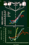
The maturation stage of enamel development begins once the final tissue thickness has been laid down. Maturation includes an initial transitional pre-stage during which morphology and function of the enamel organ cells change. When this is complete, maturation proper begins. Fully functional maturation stage cells are concerned with final proteolytic degradation and removal of secretory matrix components which are replaced by tissue fluid. Crystals, initiated during the secretory stage, then grow replacing the tissue fluid. Crystals grow in both width and thickness until crystals abut each other occupying most of the tissue volume i.e. full maturation. If this is not complete at eruption, a further post eruptive maturation can occur via mineral ions from the saliva. During maturation calcium and phosphate enter the tissue to facilitate crystal growth. Whether transport is entirely active or not is unclear. Ion transport is also not unidirectional and phosphate, for example, can diffuse out again especially during transition and early maturation. Fluoride and magnesium, selectively taken up at this stage can also diffuse both in an out of the tissue. Crystal growth can be compromised by excessive fluoride and by ingress of other exogenous molecules such as albumin and tetracycline. This may be exacerbated by the relatively long duration of this stage, 10 days or so in a rat incisor and up to several years in human teeth rendering this stage particularly vulnerable to ingress of foreign materials, incompletely mature enamel being the result.
Keywords: Porosity; enamel dysplasias; enamel maturation; matrix protein loss; uptake of foreign materials.
Figures


Similar articles
-
The chemistry of enamel development.Int J Dev Biol. 1995 Feb;39(1):145-52. Int J Dev Biol. 1995. PMID: 7626401 Review.
-
Dental fluorosis: chemistry and biology.Crit Rev Oral Biol Med. 2002;13(2):155-70. doi: 10.1177/154411130201300206. Crit Rev Oral Biol Med. 2002. PMID: 12097358 Review.
-
Mineral acquisition rates in developing enamel on maxillary and mandibular incisors of rats and mice: implications to extracellular acid loading as apatite crystals mature.J Bone Miner Res. 2005 Feb;20(2):240-9. doi: 10.1359/JBMR.041002. Epub 2004 Oct 11. J Bone Miner Res. 2005. PMID: 15647818
-
Crystal growth in dental enamel: the role of amelogenins and albumin.Adv Dent Res. 1996 Nov;10(2):173-9; discussion 179-80. doi: 10.1177/08959374960100020901. Adv Dent Res. 1996. PMID: 9206334
-
Time-related changes of developing enamel crystals after exposure to the tissue fluid in vivo: analysis of a subcutaneously implanted rat incisor.Arch Histol Cytol. 2000 May;63(2):169-79. doi: 10.1679/aohc.63.169. Arch Histol Cytol. 2000. PMID: 10885453
Cited by
-
Comparative Metabolomics Reveals the Microenvironment of Common T-Helper Cells and Differential Immune Cells Linked to Unique Periapical Lesions.Front Immunol. 2021 Sep 3;12:707267. doi: 10.3389/fimmu.2021.707267. eCollection 2021. Front Immunol. 2021. PMID: 34539639 Free PMC article.
-
Assessing human weaning practices with calcium isotopes in tooth enamel.Proc Natl Acad Sci U S A. 2017 Jun 13;114(24):6268-6273. doi: 10.1073/pnas.1704412114. Epub 2017 May 30. Proc Natl Acad Sci U S A. 2017. PMID: 28559355 Free PMC article.
-
The Distribution and Biogenic Origins of Zinc in the Mineralised Tooth Tissues of Modern and Fossil Hominoids: Implications for Life History, Diet and Taphonomy.Biology (Basel). 2023 Nov 21;12(12):1455. doi: 10.3390/biology12121455. Biology (Basel). 2023. PMID: 38132281 Free PMC article.
-
Proteomic Analyses Discern the Developmental Inclusion of Albumin in Pig Enamel: A New Model for Human Enamel Hypomineralization.Int J Mol Sci. 2023 Oct 25;24(21):15577. doi: 10.3390/ijms242115577. Int J Mol Sci. 2023. PMID: 37958567 Free PMC article.
-
Natal factors affecting developmental defects of enamel in preterm infants: a prospective cohort study.Sci Rep. 2024 Jan 24;14(1):2089. doi: 10.1038/s41598-024-52525-2. Sci Rep. 2024. PMID: 38267499 Free PMC article.
References
-
- Boyde A. (1989). Enamel, in Handbook of Microscopic Anatomy, Vol. V/6: Teeth, eds Oksche A., Vollrath L. (Berlin: Springer-Verlag; ), 309–473
-
- Brookes S. J., Robinson C., Kirkham J., Bonass W. A. (1995). Biochemistry and molecular biology of amelogenin proteins of developing dental enamel. Arch. Oral Biol. 40, 1–14 - PubMed
-
- Damkier H. H., Josephsen K., Takano Y., Zahn D., Fejerskov O., Frische S. (2014). Fluctuations in surface pH of maturing rat incisor enamel are a result of cycles of H+-secretion by ameloblasts and variations in enamel buffer characteristics. Bone 60, 227–234 10.1016/j.bone.2013.12.018 - DOI - PubMed
Publication types
LinkOut - more resources
Full Text Sources
Other Literature Sources

