Review
. 2014 Oct 7:5:511.
doi: 10.3389/fpls.2014.00511. eCollection 2014.

The role of dynamic instability in microtubule organization

Affiliations
Review

The role of dynamic instability in microtubule organization

Tetsuya Horio et al. Front Plant Sci. .

Abstract

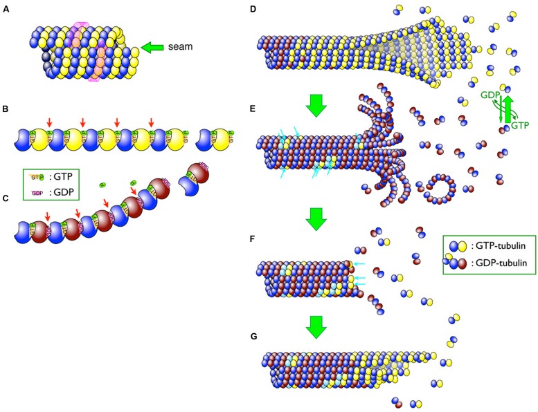
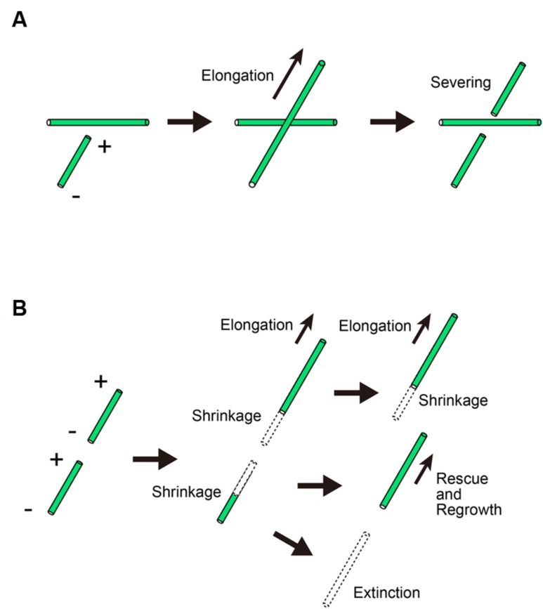
Microtubules are one of the three major cytoskeletal components in eukaryotic cells. Heterodimers composed of GTP-bound α- and β-tubulin molecules polymerize to form microtubule protofilaments, which associate laterally to form a hollow microtubule. Tubulin has GTPase activity and the GTP molecules associated with β-tubulin molecules are hydrolyzed shortly after being incorporated into the polymerizing microtubules. GTP hydrolysis alters the conformation of the tubulin molecules and drives the dynamic behavior of microtubules. Periods of rapid microtubule polymerization alternate with periods of shrinkage in a process known as dynamic instability. In plants, dynamic instability plays a key role in determining the organization of microtubules into arrays, and these arrays vary throughout the cell cycle. In this review, we describe the mechanisms that regulate microtubule dynamics and underlie dynamic instability, and discuss how dynamic instability may shape microtubule organization in plant cells.

Keywords: GTP hydrolysis; cortical array; dynamic instability; microtubule; phragmoplast; tubulin.


Figures

FIGURE 1
FIGURE 1
Schematic diagram of microtubules. (A) Lateral arrangement of tubulin dimers in the microtubule. The tubulin molecules are laterally aligned along the three-start left-handed helix (indicated by a pink wrap-around arrow) and different tubulin molecules thus meet at a seam (indicated by a green arrow). (B,C) GTP hydrolysis causes a conformational change in protofilaments. Whereas GTP–tubulins (B) have a straight conformation that fits well in the wall of the microtubule, GDP-tubulins (C) tend to curve outward from the microtubule lattice. β-tubulins bound to GDP are shaded brown, whereas those bound to GTP are yellow. Changes in inter-dimer interactions are indicated by arrows. (D–G) Depolymerization and rescue of microtubules. While newly incorporated tubulin dimers are all GTP–tubulins (D), GTP molecules are relatively quickly hydrolyzed and most of the dimers in the microtubule become GDP-tubulin (shaded in brown; D) Remnant GTP–tubulins in the microtubule are indicated by blue arrows (E). Depolymerization can be stopped by remnant GTP–tubulins as a “rescue” action (F). The addition of new GTP-tubulins (blue arrows in F) resumes the growth, and the addition of the tubulin dimers continues (G).
FIGURE 2
FIGURE 2
Microtubule behavior after crossing in the cortical array. (A) When a microtubule crosses an existing microtubule, the crossing microtubule but not the existing microtubule is severed. + and – denote the plus and minus ends of a microtubule, respectively. (B) The severed microtubule depolymerizes at the newly created ends. In some cases, the plus end starts to grow by a rescue event.

Similar articles

Cited by

References

    1. Al-Bassam J., Chang F. (2011). Regulation of microtubule dynamics by TOG-domain proteins XMAP215/Dis1 and CLASP. Trends Cell Biol. 21 604–614 10.1016/j.tcb.2011.06.007 - DOI - PMC - PubMed
    1. Al-Bassam J., Kim H., Brouhard G., van Oijen A., Harrison S. C., Chang F. (2010). CLASP promotes microtubule rescue by recruiting tubulin dimers to the microtubule. Dev. Cell 19 245–258 10.1016/j.devcel.2010.07.016 - DOI - PMC - PubMed
    1. Allard J. F., Wasteneys G. O., Cytrynbaum E. N. (2010). Mechanisms of self-organization of cortical microtubules in plants revealed by computational simulations. Mol. Biol. Cell 2 278–286 10.1091/mbc.E09-07-0579 - DOI - PMC - PubMed
    1. Ambrose C., Allard J. F., Cytrynbaum E. N., Wasteneys G. O. (2011). A CLASP-modulated cell edge barrier mechanism drives cell-wide cortical microtubule organization in Arabidopsis. Nat. Commun. 2:430 10.1038/ncomms1444 - DOI - PMC - PubMed
    1. Austin J. R., Segui-Simarro J. M., Staehelin L. A. (2005). Quantitative analysis of changes in spatial distribution and plus-end geometry of microtubules involved in plant-cell cytokinesis. J. Cell Sci. 118 3895–3903 10.1242/jcs.02512 - DOI - PubMed

LinkOut - more resources